Two DNA sequencing methods from 1977: Maxam-Gilbert uses radioactive labeling and chemical cleavage to read sequences from gel bands, but requires hazardous chemicals and low throughput. Sanger's chain-termination method became dominant by using dideoxynucleotides to stop DNA synthesis at each base, eventually adapted to fluorescent labeling and still used in modern labs.
Chapters
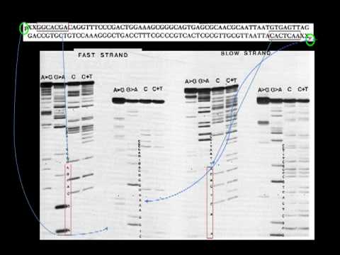
Today we will be discussing DNA sequencing, talking about 2 papers. The first is A New Method for Sequencing DNA by Maxam and Gilbert. The second is DNA Sequencing with Chain-Terminating Inhibitors by Sanger, Nicklen, and Coulson, narrated by Yan He and Courtney Morris-Gordon. Several discoveries were made that made possible DNA sequencing. The first was in 1953 by Watson and Crick when they discovered the structure of DNA, which will be discussed in another topic in this series. In 1972, recombinant DNA technology was developed which permitted the isolation of DNA fragments. In 1975, the first genome was sequenced of a bacteriophage using the plus-minus method. In 1977, Sanger and Gilbert published 2 independent methods for sequencing. They both received the Nobel Prize for this work in 1980. The base-specific cleavage method was the method discovered by Gilbert— the Gilbert group. It starts with a DNA molecule that is labeled at one end with radioactive P-32. The DNA strand is then randomly cleaved by reactions that specifically fragment its sugar-phosphate backbone only where certain bases have been chemically removed. Purines are methylated by dimethyl sulfate at the base's glycidic bond. Methylated glycidic bond of adenine is less stable than guanine. Using an alkaline treatment, this releases adenine preferentially. When a gentle acid treatment is done, it releases guanine preferentially. Pyrimidines are attacked using hydrazine, which attacks across the C4 and C6 atoms to open the base's ring. High salt concentrations target cytosine preferentially. A similar reaction is performed at lower salt concentrations, which targets both pyrimidines. Redundant information gives more accuracy and allows for less misinterpretation in this method. In the Gilbert method, a fragment of DNA was isolated. The DNA fragment is labeled on both strands with radiolabeled P-32. The strands were then separated by gel electrophoresis to get a slow and fast strand. The strands were then cut and cleaned from the gel to be used in the 4 reaction types specific— reaction types specific for each base. Aliquots of each reaction are loaded onto an acrylamide gel. The sequences can then be determined by reading the bands on the gel and determining the corresponding base. Each column is specific for a base. A strong band in column 1 and a weak band in column 2 represent adenine. A strong band in column 2 with a weak band in column 1 represents guanine. A band in both columns 3 and 4 represents a cytosine. Lastly, a band only in column 4 represents thymine. A summary of the Gilbert method is that this method is based on chemical modification of DNA and its subsequent cleavage at specific bases. The chemical sequencing method was published 2 years after the Sanger plus-minus method and rapidly became more popular. Advantages of this method is that it uses purified DNA directly, while the initial, initial Sanger method required that each read start be cloned for production of single-stranded DNA. Disadvantages of this method are that it is technically complex and uses hazardous chemicals. It's also difficult to use with high-throughput applications.
The classical chain termination MISA, or Sanger MISA, requires a single-stranded DNA template, The DNA polymerase radioactively off fluorescently labeled nucleotides and modified nucleotides, which can terminate DNA strand elongation. The DNA sample is divided into 4 separate sequencing reactions containing all 4 of the standard dioxynucleotides and DNA polymerase. To each reaction is added only one of the 4 Di-DOC nucleotides, which are the chain-terminating nucleotides lacking a 3'-OH hydroxyl group required for the formation of a phosphodiester bond between 2 nucleotides. Thus, the terminating DNA strand is tensioned, resulting in DNA fragments of varying lengths. The newly synthesized and labeled DNA fragments are heat-denatured and separated by size with— by gel electrophoresis. on a denaturing polyacrylate gel with each of the 4 reactions run in one of the 4 individual lanes. The DNA bands are then visualized by autoradiography or UV light, and the DNA sequence can be directly read off the X-ray film or gel image. In the image on the right, dark bands correspond to DNA fragments of different lengths. A dark band in a lane indicates a DNA fragment that is the result of 3' termination after incorporation of a dideoxy nucleotide. The relative position of the different bands among the 4 lanes are then used to read DNA sequence from bottom to top. In his original paper, the restriction fragments A12D and A14 was as primers on the complementary strand of S174 DNA. The inhibitor used was ddGTP, ddATP, ddTTP, and AICTP. Electrophoresis was under 12% acrylate gels. The DNA sequence is written from left to right on the apparatus, beside the corresponding bands on the radioautography. With longer restriction enzyme fragments as primers, it is necessary to split them off from the newly synthesized DNA strands before the electrophoresis. This is normally done by digestion with restriction enzymes. When this technique is used, the products are cut with the restriction enzyme at a bond. The difficulty arises if there is a second restriction enzyme site close to the first one, because this will give rise to a separate pattern of bands that is superimposed on the normal ones, making interpretation impossible. One way in which this can be avoided is by the single-site ribosubstitution method. So the Sanger method, for the summary of Sanger method, when the method was first published, sequence of about 15 to 20 base pairs from the primer site was possible. The disadvantage of this method is band pileups caused by the base pair loops, and at the time it was hard to obtain all the ddNTPs. And the advantage of The advantage of this method is it requires no preliminary detection and uses commercially available DNA polymerases and less artifacts than other methods.
To conclude, over time the Sanger method became increasingly popular due to its broad applications and ease of use. The basic concepts of this method are now used in most current sequencing facilities with fluorescent labels in the place of the previous radiolabels.