Breast Implant Health Risks No One is Talking About

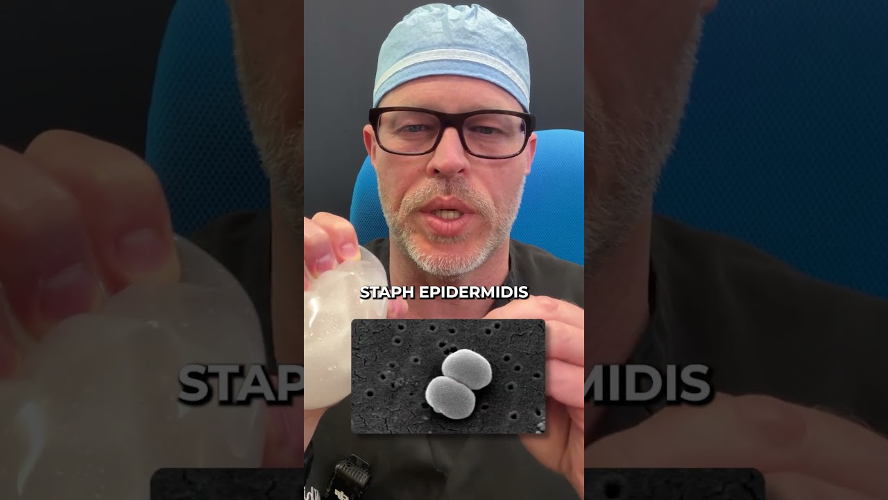
Dr. Robert Whitfield and Dr. Monisha Benote examine what happens inside the capsule around breast implants. Biofilm bacteria like Staphylococcus epidermidis can trigger chronic inflammation. BIA-ALCL, a rare lymphoma linked to textured implants, takes about 10 years to develop and shows up as CD30+ T-cells in the capsule tissue.
Chapters
Important for women to realize that when you're having these cosmetic surgeries, any kind of cosmetic surgery you're having, how your body might react to it might be different than somebody else's. And that's kind of based off of your whole body's immune system. You know, if there is the idea of infectious process that should really be cultured and sent separately because what happens is the tissue actually gets put in formalin and formalin kind of kills off everything, so we wouldn't be able to culture it after the fact. What do cytokines do? They're basically chemicals that are a response to inflammation. And when they're constantly doing that because you have this constant foreign body in there that leads to greater systemic inflammation, chronic low grade inflammation, all the way to oxidative stress and even mitochondrial dysfunction.
Welcome to the show. Today I have Dr. Monisha Bonote. She's a best selling author of Anatomy of Wellbeing and distinguished authority in plant based gut health, cellular health. Longev. With contemporal Medical Board certification, she leads in integrative medicine, functional culinary medicine, cytopathology and anatomic clinical pathology. As the founder of Walkula, she merges ancient wisdom with mind body science to forge a transformative path toward optimal health and longevity. Dr. Benode is a graduate of NYU Winthrop University Hospital Cornell and the Andrew Weil center for Integrative Medicine. She is featured in Shape, Reader's Digest and Worth of Stewart Living and share her insights globally through Dr. Binote.com serving on various clinical advisory boards. Her profound interest in the nexus of nutrition and the micro, microbiome, cellular health and mindfulness practices underscores her commitment. Through revolutionizing well being, she empowers individuals with sustainable science backed lifestyle changes that highlight the role of cellular health and nutrition in extending healthspan and vitality. All right, welcome Everybody. I have Dr. Benot. She's been on my shows before and we're in groups together. We kind of discussed at one of our meetings her expertise in pathology. And so for everybody listening, my background is in oncologic reconstruction. So whenever I started to do explants, I was taught a very specific way on an explanted specimen because obviously I learned on cancer patients all of the materials, which is the scar tissue, the skin, whatever is associated with the specimen we're sending would be sent to the pathologist for review to rule out any evidence of recurrent cancer, which is paramount. And then obviously we sent other specimens to be looked at for bacteria or fungi or mycobacterium or things like that. Now, I have had a handful of patients have breast cancer, which can obviously be found at any time throughout women's life when I'm taking care of them and doing any of these surgeries. So those would be characterized on pathology according to the standards set for examination of those tissue samples of breast. And then I have had a patient who had a B cell lymphoma, which is extremely uncommon. I think it was only the eighth in the world when we did that. And that was characterized a certain way that we talk about. I'm sure you'll find it interesting. But nonetheless, I send each and every sample off for pathology examination. So welcome to the show, Dr. Badot. And maybe you can dive into, like, what it means when we send things off her path.
Yeah, absolutely. Thank you, Rob, for having me back today. To kind of delve into something that most people don't really think about is any part of your body that is leaving you from a tissue sample to a blood sample to body fluids. It is going to a pathology lab to be diagnosed. What I mean by that is a pathologist are really doctors. Doctors were the ones with the answers to what is going on in this tissue. When we're thinking of breast. From the oncologic standpoint. Yes. You know, breast tissue, whether that's a coral biopsy, whether that's a lumpectomy, all the way up to a mastectomy, does go to the pathology lab. And we examine that in great detail under the microscope, because most people don't realize there's like, 30 different types of breast cancer. So not everybody is the same. But this is also happening in the breast implant world. When a implant fails, that implant is sent to the pathology lab. One, we're looking for rupture. Two, we're looking for what is actually growing around the implant. And additionally, we get something called a fibrous capsule. And that capsule is deeply examined under the microscope to say, all right, what's going on in the capsule? Is there? Some of the more common things we see are dystrophic calcifications, which your body basically can respond to foreign bodies by killing off cells and making calcifications. It's. It's like a debris. We see macrophages, which is a subtype of blood cell that happens to occur when there's inflammation in the body. It's a derivative of a monocyte. A monocyte is your white blood cell, and then it turns into a macrophage. And there's a number of things that they do and then we often see, you know, dense fibrous tissue, which is made by the fibroblasts. But sometimes something else even happens, like synovial metaplasia. And when we think of synovial metaplasia, that usually happens in the joint. It does not happen in breast tissue. So the body is really making changes according to this foreign body that has been entered in it. And that's where I think it's important for women to realize that when you're having these cosmetic surgeries, any kind of cosmetic surgery you're having, how your body, body might react to it might be different than somebody else's. And that's kind of based off of your whole body's immune system.
Yeah. And thank you for that explanation so that everybody listening understands. When I'm doing cosmetic cases, which are explant cases, I don't send the actual implant 2 path. Typically what happens, and I'll give you a disclaimer. The things that go are what Dr. Minot said, the scar tissue around it. So I send that off every single time. Many patients want their devices back, and so they're cleansed and then put in a sterile bag for the patient because that's their property. In a quote unquote cancer case, like I was describing, where there was a lymphoma, I could not disengage the tissues from the device. So in that setting, and, you know, this is a case by case occurrence for, for Dr. Benot, so if you can't actually take the tissue off, and typically that's a textured device, not a smooth device, I just, I tell the patient, you know, matter of factly afterwards, like it was not even something that was going to happen. So it was sent. My concern is this, and that's typically of a malignancy. And that has to be clarified by the pathologist, not by the surgeons. So every single case, the, the scar capsule, the tissue response, that scar tissue is sent and examined so that I know, you know, what's going on from pathology standpoint. And then we send a, almost like a thumb size, thumbnail size, sorry, sample for PCR analysis. So that's what our paper was published about in terms of bacterial contamination to show us at that time, like, what's there, because obviously you can't see that. And the pathologist is not going to know that either. I mean, that's a whole separate, you know.
Yeah. And we do look for bacterial contamination, but PCR is definitely going to be more of a ability to amplify, even if there is a few Bugs there. So, you know, we'll do a pathologic examination and if we see a bacterial colony, we can just, you know, gram stain it and say, oh, there's a gram positive, you know, cocci. Like if we're looking for a staphylo infection or if it's, you know, gram negative rods, so we can definitely look at that. But I do do think that all, you know, if there is the idea of a infectious process that that should really be cultured and sent separately because what happens is the tissue actually gets put in formalin and formalin kind of kills off everything. So we wouldn't be able to culture it after the fact, which is why doing it up front, you know, when, when surgeons are in there and then they're kind of like, oh, let me sen this kind of culture and let me do this. That, that's the best time to really do that. So yes, absolutely. Yeah.
I, you know, I just. Because a lot of times people like, they'll discuss with surgeons like have preferences. Right, of course. And they make this a, we'll just say an option. And so if you're coming to visit us, I don't ever feel like that's a good idea because as you know, you're going to find a handful of people with breast cancer, dcis, all these other things are going to crop up. And if you never send anything, well, you'll 100% miss it because Dr. Binot wouldn't be able to look at it. Nobody would be able to look at it. You would not know about it. And typically if I'm taking caps for material out, these are deep lying cancers that are found. Typically there are some extenuating circumstances in patients who've had really, really significant calcification responses which happen that Dr. Benot can talk about. She talked about dystrophic calcification on path. But I mean some of these things are like hard as rock that you encounter. For various reasons. People form heterotopic ossification all over the body. But these are particularly odd in the breast and they can cause a lot of pain and all sorts of things and really mask ruptures and cancers and all these things and make it really complicated for us to kind of not just plan for but manage depending on the patients.
Yeah, so absolutely, that is definitely one of the challenges when it comes to having implants is it's very difficult to do adequate imaging to see what is going on. And that's, that's where really the tissue sample comes in. But the dystrophic calcifications will mask, you know, both benign things and cancer. And I've seen it time and time again where somebody has a calcification that does show up on imaging and then they go get a biopsy. You know, usually when we're doing biopsies on individuals that have implants, it's like an MRI guided, because it's got to be so precise that we don't rupture the implant. But even that, there's only so much sample you take out with a needle that things can get missed. So this is where it's really critical to know, all right, what is going on in your body. And keeping in mind, if you, if you do have an implant and you choose to have an implant, that you're going to have to be even more on top of it to really go. Because considering one in eight women now has breast cancer, if you've got any kind of breast tissue, whatever little you have, you know, you're still exposed and at risk for this. So I think it's very critical to, to understand, you know, that process of what's going on. And then the other thing is, as you mentioned, you've seen a rare case of this anaplastic large cell lymphoma, which happens in the capsule of very rare cases of individuals who have implants. And, and it does take about 10 years to develop. So if you just got an implant, you might not, like, even. Even think about it because it really hasn't been long enough. And it was occurring with the textured implants, which hopefully now they aren't being used. But I do see women who are in their 70s, and they're like, oh, you know, like the implant has become such a part of their life that they haven't even thought about it. And they're wondering, why are they getting crazy symptoms? What's going on in their body, you know, and their doctor is trying to, like, address some autoimmune disease and it doesn't get better. And because the implant becomes such a part of your life, you kind of forget that that was actually a surgical procedure. In fact, I've even seen people not even write it down on their. Their history evaluation, because when they're thinking of surgery, they're thinking of something like not cosmetic. And I think it's very important to put all your cosmetic procedures down. You know, whether you're doing fillers in the face or implants, it's. It's critical to know because these are foreign bodies that can move around in the body.
Yeah, it is Interesting. You know, obviously many of these things are done early in. In life. There's too big. There's two big incidents where people would consider enhancement, typically early. You know, we'll say 18 to 25, and then secondarily in the. We'll say early 30s. And that's. I'm just. Those are the general, you know, don't, don't send me a bunch of hate dms. Those are generally what's going on. And I just was interviewed by somebody in Turkey, and they were asking me like, well, nobody ever spoke to me about a fat transfer or anything like that because I use those frequently have since 2004. And so you may or may not get an entire discussion if that surgeon doesn't have experience with those techniques or maybe enough to be able to discuss the risk benefits. So if someone's not familiar with or hasn't executed those successfully or has enough experience, it's going to be challenging for them to have a discussion with anybody. But because we did them so frequently with cancer patients, you know, of course I had all this different experience over time because we would do D flap reconstructions and then do autologous augmentation with the patient's own fat secondarily. So I had all this, you know, experience that's hugely important built up over time. And so if a cosmetic patient came and said, hey, you know, I. I want an augmentation. Like, if they had a really a threshold for just to go up a cup size, then that would always be an autologous fat transfer in my head. Because the risk benefit ratio of having a device is so much more significant. You would have to then say, all right, this is not a lifetime device. This is going to need to be potentially exchanged. It can rupture, it can leak and do all these things. It can get infected because any device you ever put in anybody can get infected. I mean, this is, none of this is like, complicated in terms of understanding of anybody who went to, you know, and was trained. Every implant can get infected.
And I, and I think people don't think about it because some of these surgeries, you know, we're talking about breast, but I'm thinking about knee surgeries and hip surgeries and dental implants. It's almost like an everyday occurrence. So people don't realize that, hey, I got something put in my body. It's a foreign body. And now I'm going to get a foreign body type reaction, which is basically these multinucleated giant cells and macrophages that come and identify this Foreign body. And they're like, oh, this is not supposed to be here. So what are they doing? They're secreting cytokines. What do cytokines do? They, they're basically chemicals that are a response to inflammation. And when they're constantly doing that because you have this constant foreign body in there, that leads to greater systemic inflammation, chronic low grade inflammation, all the way to oxidative stress and even mitochondrial dysfunction of this long term implant or device being in your body, which, you know, for me I'm finding it kind of interesting to see, you know, a lot of people are putting like these little like devices, like neurologic things and things in their body. And I'm like, is, does the benefit outweigh what you're risking here?
Oh yeah, sure. And much to your point, now people, you know, used to take little shots at me because I would, I'd have this hypothesis that the device like you're, and you know this because you've seen this on path, you see the chronic inflammatory cells under the scope. So Dr. Binot, for everybody listening, she can look at and she'll show some examples of these cells here in a little bit. But what she's telling you is the hypothesis that happens with chronic inflammatory changes in tissues. And now we know through science done by Dr. Muthoon Sina that the interaction between the bacterial contaminant on a device through the patient's scar capsule with the oleic acid in the breast tissue can oxidize, just like she's saying, and it forms a oxylipin, which is Oxylipin 10 home. He's also profiled other oxylipids. So just to your point, I always was like, I wasn't completely under the impression that there was no interaction. I just didn't understand it because the science wasn't, wasn't there. So he's written this really eloquent paper and people have comments on how great it is about the interaction. And then what happens downstream is what you're describing. There's a polarization change in the macrophages and that leads to much of what we see clinically in terms of symptoms. So now we have an incidence, you know, Our study showed 29% incidence of bacterial contamination on PCR analysis. We have his paper showing the interaction of the, you know, staph epi. And also another culprit in biofilm is cutibacterium acnes, which I see, you know, those are the main two. I see. And so now like the little puzzle pieces, you know, are coming Together as things are catching up. And, you know, you. Maybe you can just show. This will be on YouTube. So she is going to show us some slides so we can educate us a little bit about the cells that. That she sees as concerning.
Yeah, absolutely. So let me do a little screen share. And for those of you who are just listening to this, I do recommend you kind of go to Dr. Rob's YouTube channel and check out these images, because often we only see the outside of what's going on on our body. And if we get something on our skin, we're like, oh, we got to go address it. But realize that there is deep cellular changes going on with any foreign body that you put in. So, for example, here. So. So one thing is our tissue is not quite pink and purple, okay? In fact, all of our tissue inside our body, we're all the same color. We've got like some tan, we've got some yellow, we've got some white, you know, so our tissues are all the same color. We use a special technique and staining process. This is a hematoxylin eosin stain, which basically is a pink and purple stain to help us distinguish between the connective tissue, the nuclei, and all the different features that we need to see in order to give a diagnosis. So right here, this, what you're seeing here, is a capsule of a breast implant that was removed. So it's a capsular tissue. The fibrous tissue around does have a few inflammat cells here, these little ones with little granules. These are eosinophils, a specific type of white blood cell. But the. The prominent thing here is this reaction at the top. And this is the reaction that's kind of. If there was an implant, this would be where the implant is. It's touching it. So the body, the fibrous capsule, has made a response, creating a different type of tissue. When you create a tissue that's not supposed to be there, it's called metaplasia. So it's transformed into something. And this is specifically synovial metap. So it's a specific type of cell that has developed there. We have normal synovial tissue in our joints, okay. We do not have that in our breast tissue. So this is a response we see to, you know, having a foreign body there. Here is probably a better image of that in a sense. Like here's the capsule on the outside, and we've got this synovial metaplasia where it's changed. But then we even have something called synovial Hyperplasia, where it kind of grows into the spaces, and it doesn't really serve a function in the breast tissue. There's no reason for it to be there. It's just a reactive response to this process, which at the end of the day, could affect how your, you know, how your implant feels, how it retracts, how your breast feels in general. The other thing that we see often is something called a dystrophic calcifications. Now, there's different types of calcifications in the body. Dystrophic are usually some of these, like, larger ones. Any calcification that's found on imaging is usually examined because there are certain calcifications that are associated with cancer. So if you've ever heard the word calcifications, somebody's going to want to go further. Look at that. Another thing is that we have seen is silicone leaks, right? So if the implant leaks its material, it goes into these same pink fibrous capsules, and it kind of just sits there. But it's not just sitting there, because if you look around it, there's these little deeper purple cells. And what it's doing is now you have the foreign body, and the foreign body has even a greater response because the silicone is a foreign body. So inflammation are cells that fight inflammation, like white blood cells go, and they gather there. Here's yet another example of this refractile material, which is silicone, that has leaked from somebody's implant. This one also has that synovial metaplasia. And what we're looking at is these lymphocytes. And the reason we're looking at these lymphocytes. I'm going to get to the last slide. Is one of the rarest occurrence that occurs, but it is a bad one. So we want to be careful of that. And then people don't realize if your implants are leaking. Maybe you don't realize. Maybe the first thing you experience is actually tender, enlarged lymph nodes. Because silicone can travel through, you know, the lymphatic spaces, through the blood spaces and go into your lymph nodes. So you might have enlarged axillary lymph nodes. So this is an example of a lymph node that has been infiltrated by silicone. There's some foreign body, giant cells here responding, some kind of fat necrosis. This is not what a normal lymph node would look like. Then lastly, I just want to mention is that what we were talking about earlier with Rob was the anaplastic large cell Lymphoma. What happens there is in that fibrous capsule which is demonstrated by this red line here. The and then your implant is this center part here between the capsule and the implant, a specialized type of cell lymphoma cells which are a derivative of your B cells or a type of white blood cell, they start becoming abnormal and make a tumor there which is known as anaplastic large cell B cell lymphoma. It's a non Hodgkin's type of lymphoma. And this is what we see under the microscope. And it has a specific morphology as a specific staining pattern. It's similar to other CD30 positive lymphomas, but at the end of the day it doesn't belong there. And although the etiology is not really clear, it is believed that because your immune system is over overreactive in a way that it's being stimulated by this process. So these are just some things to realize that under the level of the skin, we really don't think about what is going on in the body on this like cellular level.
So there's just a couple of things. So the patient I had had a B cell lymphoma, but breast implant associated anaplastic large cell lymphoma is actually a CD30 positive T cell lymphoma. And those characterist CD30 positive like you mentioned, it's Alka negative. The origin obviously is the capsule. And so the treatment for that really is an explant and taking it out intact, obviously not spilling it, which is in my instance of this, it was very apparent this was not normal. I mean I've done several thousand explant surgeries and you know, what is it, 20, 25? So next year I'll be have been in practice or operating for 30 years. So you know, when things are not normal. And it was incredibly dense in terms.
Of.
You know, its characteristics because many times scar tissue, although it can be fibrous, it's not so dense that it's like hard almost. And these, these were so.
Yeah. And we do that from a gross examination perspective. When I've got breast tissue in my hands, I can really tell from the gross perspective of is this normal, is this not? Because there's certain features that a lesional tissue will have. Yeah, absolutely.
Well, I think that that's a lot for the audience for sure. You know, we continue to obviously promote our program to help just with wellness so that we limit exposures and limit risks. My biggest group that I'm trying to help now are patients who are. They have a device in place, and it doesn't matter what the device is. It can be hip, knee, breast, dental. It doesn't matter to me. And I get asked routinely, can I help them. By. By programming that will maybe push off or eliminate, you know, the need for surgery. And I. I give everybody the same disclaimer. You know, it's a function of our genetics and how we detoxify our exposures and all of our, you know, as we know, look at health parameters, you know, gut health and hormonal balance and metabolism issues. And so I. I feel like. Think we're going to help more people by expanding, you know, sharp and creating a environment for patients to not necessarily come in and have surgery, but those who do obviously will continue to send everything off and have it looked at by pathology, like Dr. Binette has described, and continue to look at other options, not just with pcr, but other ways to learn about what's going on with these patients.
Yeah, I think at the end of the day, just from our environmental influences, that some form of detoxification is key. Whether you have a device or not, the body really needs that kind of clearing. So you can restore your cellular health, your mitochondrial health, and kind of reverse some of these symptoms that somebody might be experiencing.
Absolutely. Well, thank you for coming on the show. It's been great having you again. I know you're busy, I appreciate. And so make sure everybody let them know where to find you and follow you.
Yeah, absolutely. So, on social media, I'm Dr. Benot, and my website is Dr. Benote.com. you can find me on Instagram, YouTube, Facebook, LinkedIn, all the places that if you want to go and learn.
Thank you for coming on. I appreciate it.
Thank you for having me again, Rob.
All right.