Breast Implant Removal Surgery

Dr. Robert Whitfield walks through explant surgery: why surgeons remove the capsule around implants, how they access it through an incision under the breast, and pain management with intercostal blocks. The procedure takes about 2.5 hours.
Welcome to the holistic and scientific podcast with board Certified plastic surgeon Dr. Robert Whitfield, Austin's natural choice for plastic surgery and the expert in smart laser and energy treatments.
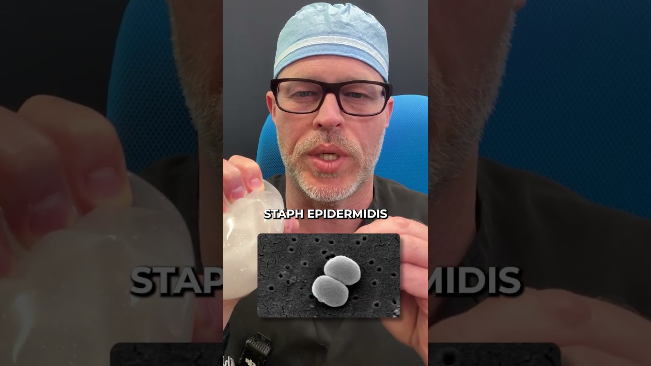
Today we're talking about explant surgery or breast implant removal surgery. When I discuss this, I try to be very specific about the goals, the goals of this procedure. In my mind, I treat everybody like they're a cancer patient. And there's a specific reason for that. One of my clients had a lymphoma. She had a texture device, and texture devices are more commonly associated with anaplastic large cell lymphoma. She did not have a seroma, which is a fluid collection, neither on CAT scan, nor mri, nor ultrasound. But it was very suspicious. It was very contracted, very firm with the lymphomas. Anaplastic large cell lymphoma. In my patient, the B cell lymphoma, the cancer cells are lining the patient's capsule that's up against the device's surface, the shell, because the irritation of the shell is what creates the activation of the lymphocytes and then creates the lymphoma. I approach this case basically like I approach every case. I'm not going to put anybody in jeopardy. But doing a more aggressive procedure than necessary, I will not compromise the care of a patient because I inadequately excised a capsule. If the capsule which harbors the lymphoma in this case, which touches the implant, is opened and exposed and spilled, or parts are left behind, as evidenced by my FDA testimony in 2019 where I testified publicly behind a woman who had had retained capsule and had anaplastic large cell lymphoma on her ribs, which is devouring her ribs because it was left there. Stories like this, experiences with patients like mine, over a decade of cancer care or breast cancer patients leave me in a unique, I guess, position. I have a lot of conservatism in me when I approach surgery. I want to do the best operation all the time. I never wanted to be the person who misses a cancer. I never want to be a person who exposes someone to more risk. Over 5,000 breast procedures, well over 500 explants at this point. I know those risks. I feel very confident I can take these out each and every time. And whether we take it out and say it's a total capsulectomy, a precise total capsulectomy, an en bloc capsulectomy, I think the goals are still the same, to take everything out as intact and undisturbed as possible for me so that I don't compromise a patient's care. I check the boxes for patients. I don't check the boxes for other doctors or entities or industry. And to me, clarifying their pathology at the end of the case requires all that material, to the best of my ability, be intact. So I remove it all as best my ability, intact all the time to give to the pathologist so they can make a diagnosis with or without staining, with, without additional information to help the patients. So there's no long term health consequences from this. It's pretty important to identify these, especially in my patient who has one of the rarest lymphomas ever found in the world. There's only eight of them. When I submitted her case to MD Anderson and for all intents and purposes, she's cured because of the way the procedure was done. Now, she didn't have anaplastic large cell lymphoma. She had a different lymphoma, she didn't have a seroma. But had I just cracked this open and took it out piecemeal, I would have compromised her care and exposed her to unnecessary risk by doing it that way. The most common way to approach this is through the fold under the breast and primary fold. That's usually where silicone gel devices are placed. Because you need a wider incision. It's more commonly done that way. You start by making the incision, removing any old scar tissue if that needs to be done, especially if it's a widened scar. Then you'll see the subcutaneous fat, which is the layer just below the skin. That's the fatty layer, which becomes important later when we talk about fat transfers, where you put the fat so we open the fatty layer and then you're going to come in contact either with the device's capsule or muscle. Typically if it's behind the muscle, if it's above the muscle, then you're going to come into contact pretty quickly with the capsule device. There's typically not a lot of tissue between the bottom of the breast and the implant capsule, which is your scar tissue around the implant surface. We'll just call the implant surface the shell and your tissue the capsule. So there are two distinct entities. Then you can easily start to see, especially if it's slightly thicker, like a tissue paper is too thin, but like a piece of paper you would write a letter on, if it's dense like that and you have some tensile strength to it, you start to gently work around it and reuse cauteri. So cautery uses current and that separates tissue it's not like the old days where we're just cutting with a cold knife, like you would slice through orange or something. That creates more bleeding cautery, causes coagulation or stops bleeding at the same time. So if you're using it properly, you're not going to hurt the actual implant capsule. You're not going to expose the implant shell, but you're going to separate tissues from and not cause bleeding at the same time. So you can what we call circumferentially dissect and you're going over the surfaces. But it all boils down to where the rubber meets the road, is how do you get this implant capsule off the surface of ribs, off the muscle? Think of it like this, towards the sternum or breastplate, however you want to term that. These are areas which repetition matters, Technique matters. Using instruments that help you matter, we use something called a freer elevator that gently raises this tissue off of the surface of the cartilage and the ribs. Cartilage is what connects the sternum to the rib. The rib is actually the bony portion. As you're raising these, there's clear planes between the layers. And so there's some pitfalls and things to avoid, like don't point instruments downward because they'll go towards the ribs. You want to stay level or parallel to the rib surfaces so you're not tipping down. That'll avoid going into the. And injuring the thorax, which contains the. The lung, of course. And a pneumothorax is what's written about a lot and described as a really bad complication from this that rarely happens. But actually, anything that's happened in surgery over time, if you've trained enough, done enough, you've seen these complications, you know how to manage them when getting that tissue off, especially in the lower inner quadrants. So towards the breastplate or the sternum, there's. Think of the breast as a pie chart, and this is the lower inner portion of the pie chart. As you come up those rib spaces, you're working your way around to the upper inner. And then when you get about halfway to the. We'll call it the border of it in the midline, you're going to transition and you're going to see muscle on the chest wall next to the ribs. And this is the pectoralis minor. And once you see that, that landmark, you can choose to keep going or you can switch and go from the armpit towards the midline. And so why. Why does it matter? So for me, it's a Level of comfort. When I see that, I know that I'm on muscle. Now there's absolutely pretty straightforward. From here we'll take it off the muscle and be be done. Now people get stuck at this position a lot and this is why people don't like doing this because it's adherent to the rib surfaces. And so one way to go about it is start the other direction and come from the armpit where there's always muscle and just roll it from, we call it lateral to medial, but from the armpit towards the sternum towards the midline. And you're gently dissecting this with your cautery off until you get to the ribs again. And then you start using a freer elevator, which looks to me, I always call it like a little butter knife because it's round at the edge and dull and you're just gently nudging it. This is what takes time and patience to do to remove this tissue. So say for instance, like my patient I described in one of my previous comments, the patient had a cancer found on pathology. You don't know who has cancer typically. That's typically a pathologic finding in this case after the fact, until the path report is back. You never know whether or not your patient safety is compromised or the long term health could be affected by this procedure. I'm always trying to get this material off based on those experiences, based on testifying behind somebody who had residual alcl left on their ribs. So I try not to leave those remnants. I try to bring closure to these so that everything's done safely and checks all the boxes. So once that materials off, because every rib, every piece of cartilage on the thorax has a surface that can be sected off of it. You can remove, can be done safely, you can control anything that's bleeding. But usually there's really not much. Normally now there's specific nuances and we can talk specifically about them. But that's how I tried to do a breast implant removal with all the capsular tissue intact without compromising or exposing the shell of the implant. What I just described is how I approach a straightforward patient with a history of breast augmentation who thinks they have breast implant illness. And because of their symptoms, once they're explant, there are special circumstances. So let's think of a special circumstance. Somebody who has a grade 4 capsular contracture which is a visible difference deformity, higher, lower shape change, it's painful. These are typically in, in my experience, easier for me to take care of because the firmer the shell, the less difficult it is for me to get it mobilized off of the rib cage or other surfaces. Because it's tensile strength, it's strong in that you can separate the layers more easily. Think of a book that's been open, the pages have dried after being wet. They're all stiff, all right? They're not stuck together. They're just. You move them. Now, you can separate them more easily if you use this instrument and nudge them apart versus somebody who's got a capsular contracture that's sort of tense, capsule's kind of thin. Those are ones that are a little bit more difficult because as you manipulate it, it can break, it can tear, gel can leak out if it's ruptured. Each of them, they bring their own nuances. And clients always ask me, like Dr. Whitfield, do I need to get an MRI to know if it's ruptured? Do I need an ultrasound? Well, I do ultrasounds in my office all the time. I will tell you it's hard to determine a posterior rupture even on mri, even a small one, because when you start pushing it around and applying pressure to the device, what happens? It squishes out. But on mri, you're never ever going to see this because you're not applying pressure to the device. I mean, it can't squeeze out. So if you came with an mri, I would say that is great. You brought me your mri. I've read through it. It says you have a rupture or you don't have a rupture. If you don't have a rupture, I don't believe the MRI because I've operated on people with normal MRIs who are ruptured. If it says it has a rupture, for me, the plan is no different. I do the exact same thing because I either think you have a rupture or you could have a cancer. If you're already prepared for all those situations and you're treating everybody collectively the same way, where you're trying to do a safe procedure to maximize their long term health by removing entire areas safely and effectively. Then on the back end, when you're getting path reports back, I anticipate all mine to be negative. Clear margins if there's cancer, just like the patient I mentioned to you that had a. A lymphoma. So to date I've done over 500 of these. I have found two cancers. One was pre explant. The patient went on to have mastectomies And I did the explant with the mastectomy surgeon at the time and the reconstruction, when I used to perform reconstructions. And then the second patient who had a breast cancer was this lymphoma that we found. But the margins were completely clear because we did the procedure properly and did not violate the capsule in any way during the procedure. So I felt very good for her after the fact that we're able to complete that effectively and she has no long term health consequences for that. She doesn't require any further treatments. She's monitored annually, and I see her annually as well. There are times when I'm surprised at the level of rupture and extravasation of silicon gel, meaning the gel is leaking outside the patient's native scar tissue capsule because the shell has been compromised. Its native implant shells failed. And if they're old implants from, say the 80s, there's been such a specific tissue response, it creates a hard shell, like a really hard egg. And then it's fine, all that is blocked and protected. But in these implant shells that then 90s ish that deteriorate and the gel's really liquid, like if someone has a thin scar capsule, the patient's scar capsule, that will leak into the tissue like syrup. And that is an incredibly messy thing to deal with. And so we get disappointed when we run into those in the operating room because those are more of a challenge. There is kind of a fun way to control the gel. You take the back of a syringe, pull the plunger out, hook it to suction, and then use the open end of the syringe without the plunger to suck the gel out. You can get the gel out more easily versus trying to do it with just a suction tube alone, because you have a bigger opening using the back of a large syringe, like a 60cc syringe. So that's a way. Use three or four or five of those to get all the gel out. And then you could perform a more controlled capsulectomy without the gel leaking everywhere. Now, after I have this material out, we use a solution that changes the ph. And no virus, no bacteria, no fungus, no mycobacterium. No mold can tolerate significant ph change because whether they're unicellular or multicellular organisms, the cell will fail when you lower the PH significantly. That's the whole point. So we do this. Approximately four minutes of acidic solution, rinse it out. If we have ruptured gel, we aggressively clean the gel material out with scrub brushes. We do everything at that time to create healing surfaces that are free from active bleeding but raw so that they'll heal together. In those instances, there's no retained capsular material, so things will heal down. People ask a lot about, can I get a seroma? Do I need a drain? All the literature used to describe drainage of, say, a breast pocket or a tummy tuck initially was felt to be necessary. But as we've learned from the drainless tummy tuck, we don't need drains. Now you use suturing techniques to control it. So for the breast pocket, what do I do special? First of all, I widely undermine the infirmary fold, which is the bottom of the breast pocket. With or without a lift. I will do this because I want the breastfold to rise. In a way, we're just disrupting it. And in this process, it becomes contiguous or continuous, if you want to think of it like that, with the abdominal wall tissue. So the fluid's going to drain out on the abdominal wall, but underneath the skin and the tissues. Now, this is how I do it for fat transfers, for explants alone, explants with lifts, and I don't use drains anymore. My hard and fast rule for a drain is a really difficult to control ruptured silicon gel leak in the pocket. Because drains don't prevent infections, they don't prevent hematomas. So you'll see more people infect themselves, then you prevent infection. Because we used to drain everybody, because we felt it was necessary to help control that fluid accumulation in the pockets. And what we had more of was people infecting themselves by touching their drains but not properly cleansing their hands. So then we started putting little clear dressings over the drains. We started using antibiotic discs around the drains. And finally I just was like, let's just use literature and say that drains, when left in people, cause more infections than they prevent. If we technically can avoid the drain safely undermining the tissues, people will produce fluid three, four, five, six, seven days, and it'll be gone. It's a normal process. There's no more infectious agent because the ph of the solution we used caused a terminal or cytal situation in the pocket. How long does this surgery take? If I'm doing an explant alone, no other procedures, I will book the case for approximately two and a half hours. This is done under general anesthesia using what's called a laryngeal mask airway. That's an area that sits not down past the vocal cords, but in the back of the throat, so there's less of an instance of sore throat, but you still can have some. One of the big things I try to do is that the completion of that explant and the completion of cleansing the pocket is perform an intercostal nerve block. I am looking at every rib cartilage, every rib itself. I see all the intercostal muscles, which are the muscles that connect the ribs together. I take exparel, which is a liposomal bupivacaine, and I inject it into the areas where I operated. The point being is to block the nerves that run along the border, inferior or lower border of the rib, as well as the muscular surfaces of the pectoralis major minor, their nerves, and the serratus anterior and sometimes the top of the rectus abdominis. So if you do that consistently, side to side, case after case, you will diminish your patient's need for both Narcotic, which is great, narcotic leaks, all sorts of complications that we don't want. But you'll be up doing more. You'll feel better sooner. You won't wake up in pain. My patients don't wake up having significant discomfort because I've done all these steps already in an explant to block what will be interpreted by the patient's brain as pain. And so the next question I always get asked is what will my breasts look like afterwards? On our next episode, we'll talk about how we use fat from other parts of the body to restore the shape and volume of the breast. Please make sure you follow the show and subscribe to our BII email list. If you are out there wondering if you have the II and want to reach out, look in the show notes for links to our office and to read more about our practice.
Dr. Robert Whitfield is a board certified plastic surgeon located in Austin, Texas near 360 and Walsh Tarleton in Westlake. To learn more, go to drrobertwitfield.com or follow Dr. Rob on Instagram at drrobertwitfield Links to learn more about Dr. Rob's smart procedures and anything else mentioned on today's show are available in the Show Notes. The Holistic and Scientific Podcast is a production of the Axis the Axis IO.