Explant Surgeon Journey! The Drive to Explant Episode 1
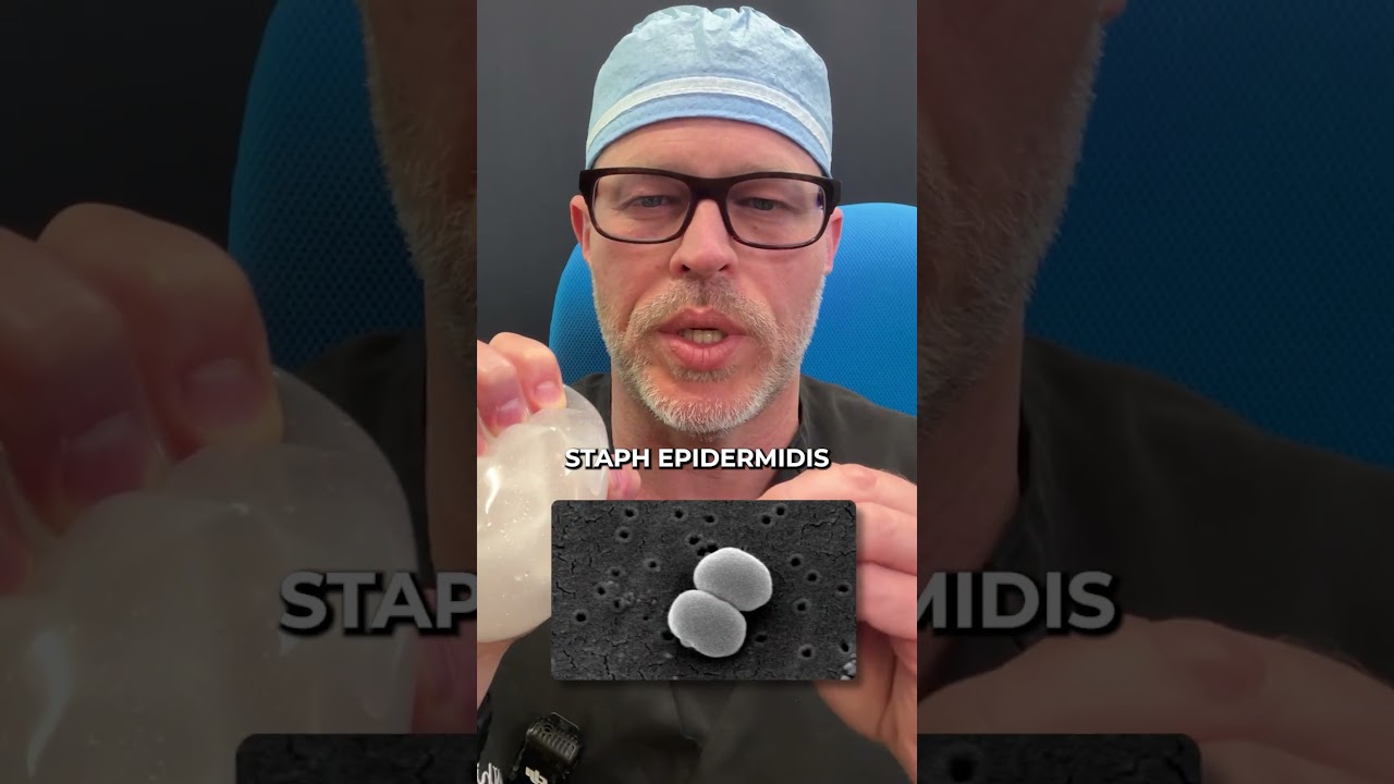
Robert Whitfield MD narrates his career path from general surgery through fellowship microsurgery to a private reconstructive practice, emphasizing breast reconstruction techniques (DIEP free flap, implant-based reconstruction, tissue expanders) and why implant reconstruction is common. He presents case vignettes (2016 explant with E. coli culture despite normal labs; 2018 explant with biofilm and negative cultures) that motivated systematic testing of explanted implants using PCR with MicroGen DNA starting Feb 2019 and reports a submitted paper estimating ~30% bacterial identification on implant surfaces.
Chapters
All right. It's a day in the life of a surgeon. Been up since 4, got a little bit of a protein drink, heading to the surgery center, and I get asked routinely on a drive to do an explant or a drive to the surgery center, how did I start doing explant? So it's a bit more of a lengthy story. I did traditional general surgery training. For those of you who don't know, that's usually five to seven years of training. It depends on your program, but it can take a year or two of laboratory work in addition to your clinical work. So I did six years of general surgery training with one year in the lab, and then I did two years of just plastic surgery. And then I chose to do an additional year of training in reconstruction using microsurgery, and then aesthetic surgery and more advanced breast surgery techniques. And then I started an academic practice and taught microsurgery for the first seven years of my career. So in the beginning of my career, it was dominated by oncology, reconstruction, trauma reconstruction. In training, it was all of the above, plus burn reconstruction. And for almost two decades, I predominantly did breast reconstruction. I also did head and neck reconstruction reconstruction and sarcoma reconstruction because breast surgery is so much more common. You know, typically one in eight women will unfortunately develop breast cancer. And the number of women that get reconstructed varies. A lot of the surgery that's done now is breast conservation surgery. So you will do a lumpectomy followed by radiation therapy, and that makes the five year survival rate equivalent to a mastectomy. So when I was training, sentinel lymph node biopsy, which is done routinely now, was developed while I was training, and that became the standard of care. So while I was training, all sorts of reconstructive and oncologic procedures were changing. They also introduced laparoscopic surgery when I was training. So taking your gallbladder out using a camera and extended length instruments became the standard of care. So a lot of things changed when I was training and first seven years of my career, I taught. I was very. I worked really hard to do my best to advance perforator flap reconstruction in the United States. That was my forte. So I did the DIEP free flap breast reconstruction, I did the ALT head and neck reconstruction, and I used the ALT free flap for lower extremity reconstruction as well. Predominantly, those were the flaps I would end up using. Those cases were lengthy and I would do several a week. I transitioned to a group private practice in Austin, Texas, in 2012, and there I did breast reconstruction. I did Head and neck reconstruction. I did all the sarcoma reconstruction in Austin. And so I was doing basically from a technical standpoint, the same types of procedures. I no longer worked in an academic setting. I no longer taught fellows or residents. It was just a private practice setting. So in 2016, I had a breast cancer patient who had a breast implant reconstruction and I had done breast implant based reconstruction as well. But because I did the IEP free flap breast reconstruction, I converted many failed breast reconstructions or reconstructions that the patients felt were painful or had capsular contracture or had injuries. Secondary radiation therapy, I would convert those to a tissue reconstruction, which is the DIEP free flap for those who don't know. The DIEP in my practice replaced what was called the TRAM flap. Because I was a microsurgeon, I could spare the blood vessels and nerves, leaving the rectus or the six pack muscle intact and then doing the reconstruction with the tissue, the lower abdominal tissue, the skin, the fat, blood vessels, leaving those intact and then connecting those underneath, really your breastbones through the blood vessels, the internal mammary artery and vein. We did that using operating room microscope. We had above a 95% success rate while doing that. And then going back to 2016, I had a patient come who wanted to have their reconstruction taken down and go flat. So the reconstructive techniques are obviously you can have no reconstruction and that would be a flat closure. The most dominant form of breast reconstruction would be the use of a tissue expander in an implant. And so I'll take a few moments to explain why that's the most dominant form. So our country is enormous, has over 330 million people in it. And once you get outside of the larger cities or urban areas, it's very hard for the hospitals to have the staffing expertise, nursing monitoring systems to take care of patients who have complex cancer reconstruction or trauma reconstruction. And that's why they get transferred to what's called the tertiary referral center. Tertiary care is the highest level of care. They will have the most intensive care capability. They'll have the most ability to provide multidisciplinary cancer care to patients. And so those are the environments normally operated in. And the final form of reconstruction would be using your own tissue. And that would vary. For over 100 years, people have used the latissimus dorsi flap. And then the tram flap became popularized in the 80s, and then in the 90s with microsurgery and more and more advancement in that perforator flap surgery became more and more mainstream. And more and more opportunities for patients to get that form of breast reconstruction developed around the United States and around the world. The first time I got really enthusiastic about was back in the early 2000s. And then I would go to meetings annually around the world to learn more about perforated flap reconstruction. So whether it was Barcelona or in India or in Taiwan, I would go to the meetings I could to get more advanced, either didactics, which was just obstruction, or hands on training in perforator flap instruction. So going back to 2016, this patient came to take down their breast reconstruction, which had been a breast implant based reconstruction. The patient on her examination had no physical findings of any problems. Sometimes you can have what's called a capture contracture, which is just firmness. And there's grades one through four that can be worsened in reconstructive cases because many times they have more surgery over time, which is typical. And then many times they're receiving additional therapies. Something like radiation therapy definitely increases the risk and rate of capture contracture. So this patient on physical exam had none of those findings. In addition, they had not been radiated. They had no physical findings, no swelling, no changes in the skin, no redness or tenderness, no pain. All their diagnostic studies really showed everything within normal limits. So the white blood cell count was normal, sed rate, CRP were normal. All the tests looking at the liver and kidney function were normal. They had nothing on any of their imaging studies over time that showed any abnormal. The only complaint the patient gave to me, which is a pretty common complaint of anybody who's had cancer treatments, especially chemotherapy, was fatigue. So with that, we went to the operating room and she wanted to actually have me take everything out intact. She actually used the term en bloc, which is a pathology term, to take everything out, basically like a tumor resection, which I had done obviously many times given my background and credentials. So I took care of her and actually was in a hospital setting because she had to be observed overnight. I did her case, took everything out. And when we're taking out tissues like that, we're trying to take the tissue out intact so that we don't. If there's an infection, spread the infection around. If there's a residual cancer or a new cancer, we don't spread the cancer around. These are just oncologic principles that we always follow. That's how I was taught. And then her surgery went extremely well. No issues with it. And subsequently on follow up at a week. Her pathology, which looks at cancer cells, suppressed cancer. Patient want to make sure she's in recurrence. Her pathology was completely normal. There was no recurrent disease, no evidence of disease, which is obviously paramount to anybod long term health. But she had on what we call microbiology exam a E. Coli infection. And so I didn't stutter. She had an E. Coli infection and it needs to be a certain amount of bacteria. So it's 10 to the sixth on a normal reported specimen. So greater than 100,000 colony forming units per high powered field. And it had a resistance pattern determined by placement of antibiotic discs when they do the cultures. And so I had no idea this patient had this problem. Obviously we spoke about it, we placed the patient on the appropriate antibiotic therapy once we knew that information, and week over week she got better. Her fatigue resolved completely. So this was a case of an underlying breast implant infection that had no physical findings and had no laboratory findings. Finance now that really bothered me. So a couple things happened. She put me on a Facebook group and let people know I did her explant surgery. So in short order, I had people calling my office to obtain explant surgery. And it was really confusing to my office at that time where I was working. They didn't understand why people were calling to get their implants removed. So that started and, you know, maybe a few times a month I would do explant procedures. And then it was Q4 of 2018, so we were doing them. I was still doing reconstructive surgery in 2018. I had a nurse come to me who really wanted to have an explant done in Austin. Stay with us sister who lived there at the time, and she was an ICU nurse from another state. And she had tons and tons of fatigue. She once again had no lab abnormalities. But I was suspicious based on her background being a nurse, obviously especially an ICU nurse, that there was potential for her to have developed a infection. And I did her explant and she had a bunch of slime around the implant. And slime, I mean, was all gooey, not like it was ruptured, but sticky like it was infected. And I went out and confidently told her family that this was infected. We just need to wait for the results and she would do better. And as soon as we knew the type of bacteria that was causing the problem, we would get her on the correct antibiotics and she would get better. Her cultures came back that she had absolutely no bacteria. And I completely disregarded this because I knew there was a bacterial biofilm. It was obvious. There's no question in my mind. So there's a research technique called pcr, which we all learned more about in the pandemic, that is a very sensitive and specific technique for identifying DNA fragments. So I developed a relationship with a research lab and now commercial laboratory Microgen DNA in Lubbock, Texas. And I've sent all of my samples there since 2019. February 2019. And that has allowed us to obtain several, well over a thousand samples. So we've done thousands of explants at this point, and we're submitting a research paper to demonstrate a 30% rate of identification of bacteria on the surface of the implant. And we feel that's the true incidence of bacterial contamination on implants. Fungus is just a handful out of several thousand. So it's really not a critical player in the contaminants. But, yeah, that's the breast implant illness story.