Prenuvo, my full-body MRI scan. Yours can save your life.
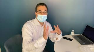
Dr. Raja Taruwala of Prenuvo explains how full-body MRI screening works and reviews Robert Scoble's scan, which found a small hiatal hernia, early kidney impairment risk, and disc bulging but no cancer or aneurysm. The scan takes about an hour and costs around $2,500.
Questions this video answers
About $2,500.
Vancouver (Canada) and Redwood City, California (near San Francisco).
Approximately every two years.
Possibly once a year.
www.prenuvo.com
So this is me, huh?
Yes, it is.
All right, well, first, the bad news or the good news? Am I going to die in a month?
The good news is things are good. That's probably the most important part of it.
And who are you?
Yes, I'm Dr. Raja Taruwala. So I'm a PhD in biomedical engineering. Then I went to med school and did both radiology and nuclear medicine as a double fellowship. So I basically combined functional imaging, anatomic imaging, as well as an engineer who basically, as engineers always do, want to make things better, quicker, and faster. And so, effectively, what we did is we basically took an mri, we tuned it to the point where it can actually go faster than almost any other MRI machine on the planet. And with it, we can scan the entire body from head to foot, and we can detect none of the 10 top cancers in stage one.
Wow.
So the goal here is to really sort of change the field of medicine. And part of the reason I built this is because. And where the name Prenugo comes from is that pre for preventative, nuvo for new. But more importantly, in the middle is Renu. And so Renu, with the nickname from my wife's childhood friend who they separated and reconnected when all of us had small kids. She had four kids under the age of five, two of them twin boys under one. And she was always tired and basically went to the doctor a few times. They pat her on the head and said, you're tired because you have four kids under the age of five. And the reality of it was she had stage four colon cancer and that'll kill her.
Yeah.
And so, unfortunately, that's not an uncommon story. And so, as a result, my friend.
Just this summer died. He was 40 with colon cancer. Right. That's one reason I was interested in meeting you.
Yeah.
And seeing what you're doing.
Yeah. And so that was kind of, you know, where you put your engineering hat on and say, wait a minute, like, how do you actually change the field of medicine for a good way? And so you basically have to start a. From a doctor's perspective, and then you take the technology and fit into what us clinicians, doctors understand, and that's what we've done. And basically, an mri, you can think of it, it's basically like a smartphone. There's all these different apps that you can build and write to make it do different things. And effectively, that's what we've done. We've actually gone and taken, I guess, a beast of a machine, very much like you can think of. An mri, like a car, and you actually get all the right parts for it to make it work, and then you write the software for it to make it go faster than anything else around.
You're the first guy that I know who's doing scans to try to prevent bad diseases. Right. And try to figure out something before it's needed. Right. Is that true? Are you the first one to use MRI in this way?
People have tried it in the past, but what they'll do is they'll basically just take a computer, conventional mri, which typically kind of focuses on individual body parts, and they'll take those little pieces, like pieces of a jigsaw puzzle, and try and put it together. And it doesn't really work well because basically what we do is we actually kind of scan in 19 different ways where we can actually look at you almost like a histology, where we're basically slicing you and looking at all the tissue and to see how. What it's composed of and how it works. And most importantly, all these MRI sequences that we use, they basically would be used in individual cards in any hospital anywhere in the world that does mri. Yeah. And that's the big difference in terms of what we do. We've just sort of solved. We've solved the speed problem. We may go way faster, but it may go even faster. You know, now we have to, like, basically get under the hood, which we now change the requirements for the fda. And so we haven't done that yet because we're starting with something that every doctor in the world understands is the best imaging modality. And we're making it work better. And then we're actually also making it so that people understand what's going on and what they're seeing. So what I've done for a decade is actually meet with every single patient and actually go through it and basically understand what they want to know. I and be able to translate this medical information technology into something that they can digest, be it good or bad. And one of the amazing things I found is no matter what information or news you give to people, good or bad, they just want to know what's happening. They just want to know what's going on.
Yeah.
Basically the most important.
It's better to know, Right?
Exactly. Yeah. And nobody's going to look after yourself better than you.
Yeah.
And your mother will scold you if you don't.
Now, the price you're charging is about $2,500. Right. To get scanned. Right. That's a lot cheaper than if I went to Xyz lab and had my whole body scanned, I'd leave. Is that true or not?
It would be because they would have to do individual pieces. And the biggest problem with doing those individual pieces is that you can't connect the dots. Right. Just doesn't work. You come together as one pack, and you don't come as a head over here and a chest over here. And so that's the biggest difference is that we connect the dots, and by doing that, we're able to see exactly what's going on and characterize the tissue. And then we have different sequences that most people don't even understand are even capable of. And that's because in the engineering or MRI physics world, we've taken things that are available and actually made them work better than anybody else has ever seen.
Got it.
Why don't we show you?
Yeah, well, yeah, let's see what I look like and keep talking about it. I visited Israel a few times, and on one of the trips, I met a company called Zebra Medical. I don't know if you know them. And they're using AI that's just come along in the last decade. Are you using AI here as well?
We are actually. We're building AI. So the whole goal of really making AI fit into the medical paradigm is to actually kind of make it more useful for doctors. You know, at the end of the day, it's like, you know, what you would want as a patient is you want to go see a doctor who's had years of experience, who can basically tell you what you have.
Yeah.
What's going to happen next and what you need to do about it.
Yeah, Right.
And so a lot of those tools, can they be built in AI? Yes, but it's very difficult to sort of come from the technology side and apply it into medicine, which is a field that collectively has trillions of years of paper literature being written. And so what we're doing is we're actually taking that and coming the exact opposite way. We're taking all the years of clinical work that makes sense to patients, doctors, and now sending it in the other direction, and we're using AI to actually accelerate that process for us.
Got it.
As opposed to trying to take AI to replace a doctor. That's not our goal. Our goal is to actually make doctors more efficient and more precise in everything we do. And so that's what we're doing. And companies like Zebra, they definitely are doing things that can be incorporated in what we're doing.
Okay. And Zebra claims that they can see some cancers On a scan like this. And you're probably using similar technology to start identifying cancer.
Yeah.
Different problems in the human body. So let's look at scan and tell me what. Tell me what you see in these things.
We'll look at these set of pictures. So it's as if you're standing looking at us.
Okay.
Then we're laying you down flat. So we'll start with this set of pictures here. So this is your right ear, this is your left ear.
Right.
And so there's your nose there in the front.
Yep.
And so then we're going through all the cerebellar tissue. And one of the key things to keep in mind is that all of these images here are clinical grade. So this is what you would see at any hospital where you got an mri.
Got it.
There's no kind of unusual, crazy sort of sets of images that nobody else knows how to interpret. So you could take these and take them to any hospital, any academic center on the planet. And the first thing they'll say is, that's not possible. And then they'll say, where did you get this?
Yeah.
Because you can't do this in like under an hour.
No.
So anyhow, it's pretty remarkable.
And being in the MRI machine is real interesting. It makes a lot of sound. You're in there for almost an hour.
Right? Just in an hour. Exactly.
And the sled moves you back and forth in this big machine, and it's making a lot of noise.
Exactly.
You might explain what is MRI for people who don't know for sure.
So MRI is actually relatively straightforward, a simple, at least in my mind. So effectively, an MRI is nothing more than hydrogen emergence. So our body has 70% water plus a lot of hydrogen on fats. And so what an MRI does, it basically takes the hydrogen or takes the hydrogen molecules or hydrogen molecules. That's normally kind of oscillating randomly. When you go into the magnet, it actually orients them all into a certain plane. And then those noises you hear is basically a temporary magnet pulling the hydrogens to an opposite direction and then letting them go back to where they normally were. And so different tissues like fats will give off signal a little faster than things like water. And so as a result, based on where you're looking in frequency, you can actually go and pick out the exact type of tissue you're looking at. So that's what an MRI is. And the beauty of it is that there's absolutely no radiation. Right. So what we're using is radio frequency, which is the Same thing that your cell phone uses, your radio uses. It's exactly the same, so it's considered harmless. Now, one of the things that you do have to watch out for with any radio frequency, just like your cell phone, is something called sar, which is a specific absorption rate of radio frequency. And so we've actually also figured out how to balance that so that you never have any issues with sar. So getting a scan is equivalent to talking about three hours on your cell phone. So it's not much at all. And if you have one of the newer, more powerful cell phones, it's walking and talking about an hour and a half on your cell phone.
Okay.
But the beauty of it is, the difference is, is that the risk of, you know, any kind of SAR or any kind of things that could potentially induce cancer is that we're now actually looking to say, is it there? Yes or no. Right. And we can answer that in a binary fashion. And, you know, we can say, for example, in your case, that there's no features of cancer anywhere that you're worried about. We got other issues.
Tell me about the other issues.
So I don't know if you want to look at this or that.
Yeah, either one.
So here we are. So, like I said, we're looking through the brain tissue. So what we're looking to see is you can see this red one, the normal gray, white tissue that surrounds the brain.
Yep. Right.
So that's. That's what we're looking for. And so we're looking to see if there's any concerning lesions based on location. So, for example, if you had a trauma at any point in your life, car accident, or fell off your bike and hit your head.
I did.
What you'd wind up seeing is actually injury to this type of an area if you hit in the front.
Okay.
And you don't have anything consuming like that at all.
Good.
Now, if you actually had things like atherosclerotic disease or hardening of the arteries and you're damaging the small blood vessels, we would see damage in this area here.
Yeah.
Also, we don't see that on you. Now, if you had things like multiple sclerosis, we would actually see those changes of multiple sclerosis in here. Again, don't say anything like that.
Okay.
So everything there, like I said, in your brain is looking quite good. Yeah. Now as well, we do these kind of pictures here where if there was something, we could tell how new it is.
Right.
So if there was something that happened recently.
Okay.
And so going back to the rest of Your brain. So now what we do is we actually go and extract the arteries from your brain. And so these are the arteries inside your skull. And so we're looking to see if there's an aneurysm. Because if there's an aneurysm and it ruptures, it's almost always fail.
Yes.
Or if we find it ahead of time, there are treatments that can. Surgical procedures that are curative.
Yeah.
And you don't have any of them.
I think my grandpa died of an aneurysm. Not in the brain, but in the aorta. Aorta, yeah.
And so you can get them anywhere in a blood vessel. And so obviously, the ones that you can't. The blood vessels you can never see, are inside your skull.
Got it.
And that's what these are. That's what bees are looking for. And so then what we do is on this set of pictures here, so this is now a side view through you. So there's your nose there in the front. And so what we're looking on these set of images for is we're acquiring these. So we can actually get a fingerprint of your brain. So we can actually see what your brain tissue looks like right now. And so we can actually compare it to you in the future, like, let's say, one, two years down the road to see if certain areas are losing brain tissue faster than they should.
Okay.
And with that, there's a lot of research in the MRI field that's quite old that can actually use it to start to predict onset of Alzheimer's before it occurs.
Okay.
And so that's.
That leads to a new question. How often do you suggest doing this?
Well, the biggest thing is, if you're looking for cancers, the only screening imaging we test. Only screening imaging test we have is mammography for breast cancer. And so that's typically done every two years based on how fast the tissue tumor would grow. So if a woman had a mammogram, and then the next day breast cancer started, two years later, it would still remain in stage one. And so therefore, we kind of suggest if you're at average risk for cancer, you know, probably every two years. Whereas if you have a strong family history of cancer or you have genetic risk of cancer, then probably more often, like, maybe once a year. Okay. So that's kind of the frequency video.
Okay.
Yeah. So then we can kind of go farther down. We can actually look at here again, your right ear and your left ear. And so what we're looking to see here is. So here's the parietal glands. So that's where there's a certain type of cancer called adenocystic carcinoma that can develop more common in men. You don't have that. And then as well in this area right here and here, that's where head and neck cancer lexus start more common in Asians. Again, you don't have that. We can see other things as well. Like if there was problems with sinusitis we would see that in there. Again, don't have that. Then we come farther down, you can see the left salivary gland, the right salivary gland. If there was a stone or something blocking it, we would be able to see those ducts dilate. You don't have that. Then we come farther down, we can actually have a look at the thyroid gland which sits right here. Again looks fine. Now the functioning of the thyroid is best checked with blood tests. So routine blood test that all doctors do. And so then I'll switch to this set of images here. And so on this set of images here we can now actually go look at the episode. So we can now actually go through and look at the chest and the lungs. So here's your right lung, your left lung. And what you'll see is you see this set of pictures here we can actually see through the lungs. And effectively what we're looking for is like a lung nodule that is cancerous on this set of pictures where basically you can think of it almost like a hardness detector. And it's very much like behave is very much like a positron permission tomography or PET scan.
Okay.
Where we can actually pick up pulmonary nodules when they are a cancerous problem. Of course we don't have any, but. And so that's good, that's good. And so then on this set of pictures here we can actually do the same thing. We're going to take you and basically rotate you around and look right through you to see if there's any concerning lesions anywhere in there. And like I said, it all looks good. And so then we'll go back to this set of pictures. Maybe I'll move, I'll move it up here. And so on this set of pictures here we're looking for any lesion in the liver. And so for example here we can see that there's that type of white spot. And you're going to see when I kind of put all these together with all of these, I can go and pinpoint exactly what this is. And you can see how I can use all these tissue Types. See that? How I go in and localize it here.
Yep.
And so I'll get rid of that. So here you can see that based on the sequence here. So pinpoint it. You can see how it shows up here, here, here, here, here. Yeah. And based on all of these different filters, as a radiologist, I know that that can be a simple cyst. Nothing but a simple cyst can be nothing else. Got it. So we know without question that's entirely benign.
Okay.
And that's one of the powers of actually having all this done in a single setting, is that we have all these tissue that allow us to characterize the tissue better than any other imaging test available. And all with no radiation. Wow.
Right.
So that's kind of what we have there.
It's amazing.
And so. So then as we go through, it.
Feels like I live in the future.
Well, that's actually what we're trying to build. I don't know if you ever see the movie Elysium, huh? Yeah. So our goal is to basically build Elysium. All right. Like, that's what we're doing. So this is like the first step of it where we can actually go and detect what's wrong and then, you know, eventually basically be able to prevent late stage disease from happening. Like, that's. That's the goal.
Got it.
And so that's what we're looking for. And so going back to you, so here's your esophagus. So we can actually go into the esophagus and we kind of see is that here it connects to the stomach and you see this little dark line right there. So it's a diaphragm. So that's where the esophagus comes. Comes right through the diaphragm to connect to the stomach. And yours is a tiny little bit high. So you have a small, little, tiny little hernia there.
Okay.
Pretty small. So with that, typically people can kind of feel like a sort of a sharp stabbing pain, particularly after you eat a lot of hot, spicy food.
Yep.
So that's.
That's. I. I can't eat hot wings. That's probably the reason why.
That's not probably. That is the reason why. That's like the best. And that's like the power of whatever I can show you. Right. What we've done, we actually have the ability to combine your symptoms with the images to the point where the images can now predict your symptoms. So instead of you trying to tell me a story about what your symptoms are, I can tell you what your Symptoms are based on the imaging, which is entirely objective. Right. So that's a hover reporter. Permeable is.
Okay.
And so now we'll keep on going. So now we can actually look at your gallbladder, which is right here. So we know that you don't have any gallstones. So there's no gallstones in that. And then as well here, we can see the spleen, which also looks fine. And then here we have the pancreas coming the whole way across. And we do all these different filters. So, for example, I'll just put these on top. So on this one, we're looking for increased I deposition in the liver, which can occur more commonly in men. And then on this set of pictures here, what we're looking for is fatty liver disease, which is a precursor for diabetes. Again, you don't have any of that. That looks good. Then I'll go back to this set of pictures. Back to this set where again, like I said, here we can see the pancreas. And so we can actually see all the detailed pancreas there. A pancreatic duct. See a little white dot? Yep. And so that allows us to make sure that everything in the pancreas looks perfectly normal. But more importantly, when we come all the way over to this set of pictures, we can actually now go and look at. So here, I'll zoom this in. So here's another view. So there's your gallbladder right in there. And so here you see this little white line, that little curved line. Yep. So that again represents the pancreatic duct until we know that your pancreas is perfectly normal. So with pancreatic cancer, one of the things that commonly happens is the duct gets blocked.
Yes.
And then that duct will start to expand. And so we can tell if that duct is expanding. We can see if there's a tumor mass. So we're able to look at that area of the pancreas in incredible detail.
Wow.
We can do that detail on every organ in the body. Wow. So that's what we're able to see. Talking too much.
Yeah. This is awesome.
I think I inhaled a piece of my mask. So then as well, I'll go back to this test.
Yep.
So here you can see. So there's your right adrenal gland. There's your left adrenal gland. Right at the top of the kidney. So here's your left kidney, your right kidney. And so we know that your kidneys here also look perfectly fine. We don't see any areas of concern there.
Okay, now I have a low create or a high creatinine level. And so my kidneys actually aren't working all that well. Yep.
And exactly as I kind of expected.
Okay.
And the reason why. Now we can go back to these two sets.
Yep.
So when we look at the. And this is where companies like Zebra are. Will be incredibly valuable is because what we can see here is when we look at the kidney. So here's your left kidney, your right kidney. And then you can kind of see here. I'll zoom this in. You can see here all around. This is like what we call the peri. Renal fat. And you can see that there's this kind of jagged little lines that we're seeing here.
Yep.
Coming off the kidney. If we go to this set of pictures here, you can see the little jagged lines are all bright white. So when we see that, that's actually quite common from what's typically called sweating of the kidneys.
Okay.
And so nobody knows exactly what causes that, but it's actually associated with two things. One, early diabetes, also associated with things like high blood pressure. But also, most importantly, those can be associated with developing early kidney failure.
Got it.
So we're actually seeing those signs. But now, interestingly, this sign has actually been around. Radiologists, we see this all the time. But it hasn't been until probably the past couple of years that somebody actually published this correlation of the findings of core kidney function with this type of imaging.
Interesting.
And conventional mri, where somebody's just kind of looking at just a little band through the abdomen. So people are publishing all this literature. And the beauty of what we're doing is instead of just looking at a little band, we can actually go and compare everything. What you would want to know is like, well, the risk of diabetes and high blood pressure, is that going to affect other organs, in particular the brain? We already know the answer to that. Which in your case, there's no.
Got it.
So that's. That's kind of, you know, where we are, what we're looking for. So then we can keep on going further. You asked about the aorta. So, you know, so here we can see the aorta here. So you can tell you that there's no abnormal dilation of the aorta. So no aneurysm in the aorta. So it looks perfectly normal.
Awesome.
It's normal in size. And so then as well go through this, we can look at a large bowel. So this here is a large bowel. And so this is where the small bowel connects to the large bowel and so we can basically go and trace across the large bowel and see if there's any concern in the lesions. And typically, what I'll do is I'll put.
Now, I just had a colonoscopy a.
Year ago that's perfect because it's the best test. Right. And the reason why it's the best test is because a polyp, if it's in there, they snip enough when they're in there, and they'll never turn into cancer. And that's what we want everyone to do.
Yeah.
Right. You know, here you can see, for example, this. This represents stool in the bowel. And so when you do a colonoscopy, as you know, the worst part is the valve prep, where they got to clean out all that stuff. And so there's no point doing anything like that for our test, because we. Or any virtual colon exam, because if there's a problem, you have to go clean up the bowel again and do a real colonoscopy. So just start with the first best test. Whereas here we can go through. We can see all the large bowel and see if there's any abnormalities or problems anywhere in there. We can basically go through and run the entire large bowel. Like I said, things look. Things look good.
Yep.
So don't see anything concerned there. And so then, as well, we can have a look at the prostate. So here you can see these are the semavesicles. See the little kind of round, bubbly things, so we know that they look perfectly fine. And so here we have the prostate gland, so we can see it. And on this set of pictures here, this is our hardness detector. So you know how doctors do a rectal exam where they put a finger in the rectum and feel along the back wall?
Yep.
Well, this is basically us doing that virtually so we can see if there's any hard spots in the prostate. And again, like I said here, it looks. Looks good.
That's awesome.
And we're seeing just a little bit of fibrosis at the front. See how it's kind of dark up in here?
Yep.
And so we know there's a little bit of fibrosis. And so at some point, you have some prostatitis that the left behind and scarring. And then one of the key things is that repetitive inflammation in any organ can lead to cancer induction.
Okay.
And so it's one of the things that in the prostate, typically, there is next to no feeling, so nobody knows when it could happen, but we can see it and so then this set of pictures. Now this is sliced right down the middle. So there's your chin there. And so here we can see the spinal cord as it's coming the whole way down. Right. And so if we go and zoom in, you're going to see. See that these represent the discs in between each of the lungs and the vertebrae.
Yep.
And so as you come across here, this represents the nerve that's coming out at every level. Right. So you can see the white is the insulating fat tissue that goes and protects the nerves. And you'll see here when we get into the lower back region, you'll see the amount of white that's insulating that nerve gets less and less.
See that? Yep.
Compare up to here. So we know that we can see that these discs are actually starting to bulge out a little bit, making that space around the nerve a little bit narrower.
Yeah.
Right. So you have. These are called deter disc.
My mom had a bad back, and so she had a. Had to have discs fused together. Exactly.
You know, and the whole thing is like, that's what we want to prevent.
Right.
And the whole idea is you want to be able to see it before it becomes a problem that requires a major surgery like that. So you can basically either delay or hopefully even prevent that onset further happening.
So what's your idea? To prevent more damage there?
So typically, like, the most important thing to prevent this. Bees from progressing is you can't reverse the clock on these. Once you're there, they're there. But the best thing to do is actually help stabilize the spine. And the way you stabilize that spine is actually improving core strength.
Okay.
So for example, here.
So you want me to do some exercise and I'm.
Yes, indeed. And basically what you're telling me is you don't want to. What I'm going to say is, like, you know, look, you can see that the writing is on the wall. Yeah. You know, if you don't, well, we will basically be watching this get worse and worse.
Got it.
You know, so the whole thing is, you know what your mother went through.
Yep.
Do you want to do that?
No.
Exactly. So the choice is yours.
Okay.
But most importantly, we could actually see this before it becomes that kind of a problem.
Got it.
So that's the whole idea of renew, basically preventing things before they progress to become a problem. And that's what it's all about. Instead of reacting to a problem once it's there, the whole idea is preventing it from becoming a problem.
That's really amazing.
So that's the pack behind what we're doing. And so then again, just to kind of give you an idea, so here we're basically kind of looking at you. So to get an idea, like the level of detail, what we're looking at. So you can zoom in, you can see all these little blood vessels underneath your skin.
Yeah.
So that's the level of detail that we have here.
Wow, that's cool.
That's you.
That's me, yeah. Wow. Well, thank you so much. So anybody can get scheduled here. I think we talked a little bit about insurance.
And I think Andrew probably knows that better than I do in terms of how it works. But basically anybody can get scanned here. And the biggest thing that we're doing that's really important is that we are coming from a clinical perspective with the idea like, how do we prevent bad things from happening to good people? And that's what we're doing. We're taking tools that every doctor understands, and we're basically enhancing what they would know. Most doctors don't even know this type of technology exists. And so as a result, there's all sorts of interesting technologies that are out there that typically come from the technology side, not the clinical side. And so there's a lot of sort of misunderstanding about what can be done. And probably one of the biggest. The biggest sort of, I guess, bad service to the field of imaging is that people were using CT or CAT scans to actually screen for people. The issue with that is that, number one, there's radiation. Radiation we know induces cancer. Number two is that you can't detail the tissues at all. You have no idea what's going on. Whereas here, as you can see, there's all these different sequences that are filters that are looking to characterize tissue in a very detailed histology way. And we have you encompassed in one go. In one go. So we can actually go and pinpoint exactly what's going on. That's where it's actually become powerful to the point where if there's an issue, you know that that's exactly what it is. Can be nothing else.
Yeah. And you have two locations, one in Vancouver and one in Redwood City, California. Exactly. Near San Francisco. Yeah.
So I started this in back in Vancouver 2009, with a hunch as an engineer that, hey, I can make these machines work better than they do. And then, you know, we kind of worked again with physicists myself, and basically a lot of scanning, and we actually started to get this to work better, better and better. And then for close to a decade, We've been doing this and, you know, like I said, at this point, I've seen like royalty, Grammy Award winners, Academy Award winners, politicians of every flavor, but most importantly, the people I kind of do, I do this for is actually for like the single mother with, you know, they are basically the nucleus of the family. And going back to renew. You lose a mother, it's a generation loss.
Yeah.
So that's what it's for.
I agree.
So the goal is to really kind of change that paradigm so that bad things happen to good people and families don't suffer as a result.
Well, I love what you're doing. It's a new look at healthcare. It's the future, and it'll be interesting to watch you put these machines to use and continue improving them. One last question. How has this field changed in the last decade? Because the technology to do this, I think probably is radically different than when you started 10 years ago, right? No, not really.
Sad, but true.
But no, the AI is changing now.
The AI is changing. And again, like, where the AI is really coming from, it's again, more the technology trying to improve. Whereas the field of medicine is kind of going, what do we need it for? We know how to diagnose stuff without AI.
Yeah, right.
And is AI going to basically going to replace us? Is sort of one of the big fears that doctors have, which is really, I kind of think, somewhat unjustified. The whole goal of where AI should be, should. How it should be used is to basically kind of make us better.
Yeah.
Make everybody better. You know, and ultimately the goal is to, like I said, make that a lease in the machine where, you know, it can go and scan through this and find problems. And then you as a radiologist can say, okay, you know, this is what's going on. You need to be, you know, go to the next step and have this treated. And so what. We know that in the developing world where, you know, people like myself as radiologists are extraordinarily expensive to train. They'll never be able to get to, like, the amount of radiologists that is present here in the United States.
Yeah.
So they're gonna. They're already, like, desperate for this stuff, like these type of tools.
So this could democratize this kind of real healthcare globally.
Right. Like, I've had sort of leaders of countries that are sitting with me in Vancouver saying, you know, here's a blank check, write what you want and come, come here, come to Asia and we'll build you anything you want to do all this stuff. And I Kind of said, you know, that doesn't seem like the right idea. You know, it's kind of like, how do we sort of improve from grassroots, patient by patient level?
Yeah.
You know, it's not all about money. It's actually about saving lives and preventing bad things from happening to good people.
Very cool. Well, thank you so much. Where do I learn more about you?
You can find more about us on the prenuvo website. So. Www.prenuvo p r e n u v o.com Very cool.
Thank you so much. And thanks for showing me myself for the first time in 55 years.
I was actually going to show you. What's also really neat is like, what's more important is it because part of the goal is actually educating people about what we do. So we've actually built this whole prenumo app.
Okay.
That allows you to basically kind of get all your information in your own.
Pocket, and so you can take it to your doctors and carry it around with you.
And even if you're traveling, like, the whole goal is no matter where you are, you actually have this. And so what we've done is we basically go. You can kind of see, we go system by system where we go through everything and describe the different body parts. And so, for example, here in the respiratory, we explain to people what it is, you know, and in understandable terms. And so then we go through, for example, here, these are all the things that we're looking at. And so here we describe, you know, for example, in this case, the sinuses. There's some sinusitis. You know, what is it? We found it. Here's a picture of what it looks like. So we've parsed through all those images to kind of show where the issue is so you understand it. And now there's all this information of. Every patient would want to know what causes sinusitis, what are the symptoms of it. And so if you go and you click on it, you'll get even more detail about every single condition that we can see so that you as a patient are educated about what's going on in your own body.
That's amazing.
And so that's the Prenuvo app. And then as well, all of these images, and they're all secure and de identified, so only you know where they are. You can basically go and you can take all these images, you can go and point and click on everything, and you can kind of see exactly what's going on anywhere, anywhere in your body. And we explain what each of these sets of images do so that you have an encyclopedia of yourself.
That's amazing.
In your pocket.
Thank you so much.
So that's. That's what Pernuvo is. This is a download of my brain for everybody.
That's awesome.
Well, thank you so much.
It's an honor meeting you, and it's an honor going through your machine and getting my scans done. I appreciate it a lot.
We'll put all this together, and then you'll actually have all this on the Preneubo app. So you have a copy of that.
Great. Thank you.
All right, pleasure.
More from Robert




45: Sandy Carter Unveils the Future of AI: Myths, Agents, and Creativity | SXSW Insights