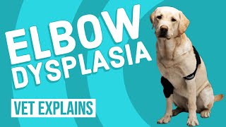
Dr. Lera Clone breaks down elbow dysplasia in dogs, a condition affecting large breeds starting at 5-14 months old. Males are about 75% more likely to develop it. Treatment ranges from arthroscopic surgery to stem cells and PRP, with most dogs recovering in 8-12 weeks if caught early.
Chapters
Hey, guys. Dr. Lehre here today. I am here with Fauda, and Fauda is here because he's been having some leg issues. His front leg. Ish. Front legs have been bothering him for probably about six months now. So we're going to get into what's called elbow dysplasia. We're going to talk about some of the different causes of it, what it actually means, how we go about diagnosing it, what are the dogs that typically get it, when do they get it, and then what are the typical treatment options and what the prognosis is. Stay tuned, and we'll get into that momentarily. All right, guys. So elbow dysplasia, there are four different things that typically make up elbow dysplasia. And it's not all different things that add up to elbow dysplasia. There are different conditions. So there is one that is called un united anconeal process. There's another one called fragmented medial coronoid process. There's another one called elbow incongruity. There is another one called osteochondrosis desiccants. Okay, so for the most part, all these. Or all these parts, all these conditions are different variations on how the elbow is jacked up. And so, for example, elbow incongruity, which is the easiest one, it just means that the joint or the bones that make up the joint on each side don't necessarily line up exactly the way they should. And so then what ends up happening is they kind of wear abnormally and they eat up the cartilage. And so that's the simplest one in terms of how it happens ununited and canal process. So that is something that typically when the dogs are growing, there's a part of the elbow that doesn't necessarily form or grow the way that it should. And so that's pretty much what the anchoneal process is, the fragmented medial coronoid process. That is something where it's not necessarily trauma induced, but there is a little piece of the elbow that ends up breaking off. And so then it's almost like this. This little piece of bone that's in. That's not almost. It is this piece of bone in the elbow joint that I usually explain to people it's kind of like a rock in your shoe all the time. And so, of course, if you think about that, that's super uncomfortable. And if you leave it in there long enough, it's going to cause damage to the bottom of your foot. Now imagine if you had something like that, but in your gum. So that's just going to tear up your gums a lot faster. And the last thing is something called osteochondrosis desiccants. We did another video on that condition specifically. And that condition is something that again is transmitted through the genetics. It is usually seen in younger dogs. And so that is something where there's a. The thought process is that there's a flap of cartilage that outgrows blood supply or there are a number of different things that kind of happen. And so then you have kind of like this like hangnail of cartilage in the joint. And it may not be as uncomfortable as per se, a piece of little bone in there, but it is still uncomfortable, still causes a lot of inflammation and tears up the cartilage. So we've talked about exactly what elbow dysplasia is. Now we'll talk about the different kinds of patients that will see it. This is something that is typically seen in large to giant breed dogs. So we're talking about new fees, we're talking about golden retrievers, we're talking about German shepherds, we're talking about chow chows, just to name a few of the breeds out there. And so it is something that is typically seen in dogs between 5 to 14 months of age, not years, months. So this is something that does happen very, very young. It is also something that, I think there's one study that says that males are 75% more likely to have it than females. So that's something else to take into consideration. Now, why did Fouda come to me? So Fouda had this particular history of elbow or lameness on, where he walks really, really funny with his legs swinging out and that kind of stuff. And he had been to another veterinarian and even a surgeon, and they did X rays on him, they did a orthopedic exam, and they weren't able to really elicit any consistent signs of pain. So the, you know, the owners still wanted to get an answer because he wasn't getting better. And they were concerned that he, that there was something that could potentially be done for him and they wanted to make sure that he had the best quality of life. So they came to us at Heron Lakes and we went ahead and did an orthopedic exam. On that particular day, I thought I was able to elicit pain on his elbow. And so obviously first thing for me was elbow dysplasia. The next thing was usually what I tell people is if there's issues with the elbow, a lot of times the best form of imaging is usually going to be a CT scan. And so the reason the CT scan is better than X rays is because you can typically magnify in there. You can see, like a 3D version and a bunch of different angles that you can't see on X rays. And so a CT scan is going to be superior. Now, when we had Fouda come in for his orthopedic exam following that with my surgical expert that particular day, Fauda was not limping. The surgeon couldn't elicit pain on any of the joints. And so we ended up doing X rays again. And so you guys would say, well, why would you do X rays again? You said that the CT scan was best. Well, the CT scan is the best, but you have to put them under general anesthesia. And it gets into the thousands of dollars for the cost of a CT scan. So if there's a possibility that could potentially go ahead and look at getting that diagnosis with just X rays, that would have been great. The other thing was that at the time when Fouda first had his X rays with the other surgeon, his growth plates were open, and at this point, his growth plates were closed. So we weren't able to see anything on the. On the X rays. So we set him up for the CT scan. The surgical expert at that time said, hey, look, you know, based on the breed or his size, his age, and the fact that he wasn't really able to elicit any sort of pain anywhere, he said, my money is going to be either on the elbow or on the shoulder. And so we did the CT scan today. The preliminary report coming back is that we think that he has issues with potentially both of his elbows, not just one, but both. And so in terms of treatment, what we do, surgery is typically going to be the choice for the majority of those different conditions that we discussed. The sooner you treat them, the better it is for the patients. Now, you say, of course you're going to tell me to do surgery sooner rather than later, because you're a veterinarian, you want to just want to run the bill up. No, what ends up happening is if you want to damage that cartilage, there is a very good chance that that cartilage is permanently damaged. And so the issue is, the sooner you get in there, the faster you get that thing out, the less damage that has happened to that joint. And so if you wait longer to do it, then more damage is building up in that joint, and then your dog's joints are going to degenerate that much faster. Now, today we do have the advantage of being able to do stem cells and prp and there's other things coming out. I'm going to do a video on a new treatment that's just come out probably within the last few months. It's a radioactive injection. I'll be really interested to go over that with you guys. But these are things. The stem cells is something that does help to repair the cartilage in that joint, and it does definitely give us the advantage of trying to give them almost new cartilage that they didn't have before, and that wasn't an option before. So we're going to wait to get the CT report back. And once we get that CT report confirming from the radiologist that indeed he does have the. The elbow dysplasia, then we'll move forward with that treatment. Now, the other thing that you'll notice is that Fouda does have some fluids on. The reason he's got these fluids on is because he had some dye go into his circulatory system when we were doing the CT scan so that we can try and highlight the different parts of the body that we're concerned about and so that we can get better images. So that's why he's on fluids. We try to make sure that we dilute all that out if indeed we do see the stuff on his elbows. The best form of treatment or surgery is going to be arthroscopic. That means using a camera to get into that joint, not actually doing an arthrotomy, which is actually opening up the joint. It's still probably going to be a pretty decent recovery process or length, so probably looking about eight to 12 weeks. And you're going to be doing some controlled exercises and that kind of stuff. On the patients that I've seen with the osteocondrosis desiccants, they took about a good 10 to 12 weeks before they resumed normal activity, and they were probably close to their peak recovery. Other than that, that's pretty much it. The prognosis is good, but you do have to do other things once that is fixed, whether it's stem cell therapy, whether it's stuff like chondroitin, glucosamine, adequan, turmeric, omega 3 fatty acids, CBD, oil, whatever it is that you can think that helps with reducing inflammation in the joints, that's what we're going to want to do. In combination with making sure that you keep your dogs as light as possible. The heavier they are, the harder it's going to be on their joints, the faster those joints are going to degrade. So please, please, please, if you guys have one of these dogs that has one of these issues, make sure that you go ahead and, and you keep them light. And you're also proactive with the supplements. Thanks for watching. Please engage with us on our social media. Really helps us and have a blessed day.