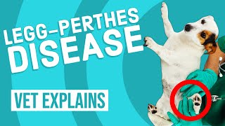
A veterinary client‑education YouTube video by Dr. Lera (Dr. Lera Clone) explaining femoral head necrosis (Legg‑Calvé‑Perthes) using a case example (Max, a Yorkie). The video defines the condition, describes typical age and breeds affected, explains exam and radiographic diagnosis, and reviews treatment choices (medical management, femoral head ostectomy (FHO), and total hip replacement) plus postoperative rehabilitation and follow‑up.
Questions this video answers
3 to 13 months (most commonly 5 to 8 months).
Bichon Frise, Yorkshire Terrier (Yorkie), West Highland White Terrier, miniature Schnauzer, miniature pinscher.
Max — 12‑year‑old male intact Yorkshire Terrier (Yorkie).
About three days.
Around four to six weeks post‑op.
Chapters
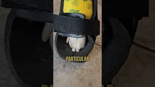
Guys, My name is Dr. Lara. Today I'm here with Max. Max is a 12 year old male intact Yorkie. And he where the. The topic of the video today is what we call femoral head necrosis or leg calf Perthes disease. So if you guys want to know what that is, stay tuned and we'll get into it momentari. All right, so leg calf Perthes disease or femoral head necrosis is typically what what it is. And let's see here. I'm gonna hand Max back to his mother, so if you just hold on for a second. It's because he wants to get down. Let's see here. Okay. She made sure to to brush him right before he got on video because she wanted to make sure that everyone knew that he was well taken care of. And he is loved, that is for sure. But in regards to femoral head necrosis, so typically what that means, let's talk a little bit about what exactly it is. The femur, which is typically this bone right here. That bone has a ball, and then that ball goes into a socket on the joint like that. And we don't know why. We thought there might be some genetics. We thought maybe there was a lack of blood supply because it is a condition that is more commonly seen in smaller breed dogs. But what typically happens is the head of the femur will go ahead and it'll start to die for one reason or another. And we think maybe there are a couple of different hypotheses out there or thoughts as to why that's happening. Again, we don't have a hard and fast reason as to why that is. And so as it dies, instead of having a nice round and smooth ball, we start to get this rough and dilapidated rock almost that is going ahead and moving around in that hip joint. And so it's tearing up the cartilage. It's causing a lot of pain. And sometimes what may happen is you may also have some hip dysplasia where the plate is a little bit more open than it should be and it causes a lot of pain. Now, typically this is something that is seen in dogs between 3 to 13 months of age. Most commonly, it's seen in dogs between 5 to 8 months of age. There is no sex predilection for the dogs that experience this. Some of the more common breeds are some of the most more commonly known breeds that get this would be a Bichon Frise, or a Bichon would be a Yorkie. They have the West Highland Terriers. You have miniature Schnauzers. You have miniature pinschers, just to name a few of those breeds. And so the way that it's going to present the dog is going to go ahead and one day, all of a sudden get up and you'll notice that they'll be either lame on a back leg or they're not going to want to use it, or you might notice some stiffness. And when you end up taking them to the veterinarian, they'll do an exam. And typically when I'm doing an orthopedic exam on the back leg, work my ways from the toes all the way up to the hip. And when I check the hip, what I have found that seems to be the part of the physical exam that gives me the clue that there's an issue is when I pull the leg back. And so when I'm pulling that leg back, it really puts strain on that hip joint. I know that this is my shoulder joint. I'm just pretend with me, okay? And, and so what ends up happening is that tells me, hey, we got pain here, not in the knee, it's in the hip. We go ahead and we shoot X rays. Now when we shoot those X rays, you're going to see that there is a round ball on one side, nice and smooth, typically in the socket. And then on the other side you're typically going to see that gnarly looking femoral head which is in there. And it's not even going to be a question. The majority of time, I normally, I want to say 99.9% of the time, send all our X rays out to a board certified veterinary radiologist to make sure that we're not missing anything. And when I saw Max's X rays, I saw, you know, it was obvious. But I still send the X rays out because there are other things that maybe could be missed because, you know, we see something so blatantly obvious. Now when we go into talking about what the options are for treatment because these patients have such severe disease, it is something that usually requires such severe disease at such a young age. It is something that requires surgical correction. Some people might say, well, I can't afford the surgery, or you know, I don't want to put them through the surgery. And you can try putting them on nonsteroidal anti inflammatories like Daramax, Prevacox Metacam, Rimadyl or Gallipram. The issue is that you're using these drugs which we will use on patients in the last quarter of their life for osteoarthritis. And it Works to a degree, but then it won't be enough. And so when you're talking about a puppy that is experiencing this degree of pain at less than a year of age, or maybe a year and a half at best, they're not going to be able to make it their whole life with just drugs. And so the, the surgical corrections that usually will be done will either be what we call a total hip replacement. And so what they do is they go ahead and they remove that femur head and they go ahead and put an implant, a rod implant with a ball. And sometimes, if I'm correct, and I may not, but they usually put a false plate in there as well, or a titanium plate or whatever metal implant it is that they put and it gives them a brand new hip. Now, that procedure is pretty intense and there are only a few places that usually will do those kinds of procedures. The level of sterility that's also required for that is super duper high because you're putting this, you know, this implant, this metal implant in, and it's in the joint. And so we really need to be super careful with those. That being said, a procedure that has been done for a very long time is something called an fho. And so what FHO stands for is femoral head osteoectomy. And so again, they go in, they remove that head, they do nothing with the socket joint. And typically what the body, what will happen is the body will go ahead and form a rubbery joint or a false joint, and the patients will then learn to use that leg with that level without any sort of ball going into the socket. Usually the muscles will go ahead and build up and provide the stability to keep the leg where it should be. This is not something that is done typically for working dogs. So if you have like a rescue dog, search and rescue dog, or a scent hound or, or a hunting dog, you're not typically going to want to do the fho. You're probably going to want to go with a total hip replacement. But those dogs, usually those larger breed dogs, are usually not going to be suffering from this particular condition. They might suffer from hip dysplasia, but they don't typically suffer, suffer from this. The other thing that you'll have to realize is there will be physical therapy involved after the procedure. One of the things that these smaller dogs a lot of times tend to do is they will go ahead and instead of using that leg that was surgically corrected, they'll say, oh no, that's too much work. I don't really want to have to deal with that. I can very easily get around with three legs, and they'll hop around on three legs. So it is crucial that you go through the physical therapy if you want to have. You know, there are places, there are veterinarians that do specialize in physical therapy alone, then there may be places like us, where we go ahead and we work. You know, we have some different protocols that we use for different kinds of surgical procedures. And the physical therapy afterwards, we typically will incorporate laser therapy into the physical therapy. So that way it helps with the recovery. The other thing that also helps with the recovery immediately the day of surgery and the first few days afterwards is something called nocita. Nocita is something that you can ask your veterinarian about. It is a local anesthetic that lasts for three days. And it's something that's game changing for these patients when they're having orthopedic procedures and even non orthopedic procedures, because the surgical site will be numb on day one, after surgery, it'll be numb on day two. And as the pain level drops, when they first start experiencing that pain level, instead of experiencing it up here, they actually start to experience it down here. And so then the drugs that we use to manage that pain can really tackle that a lot better than just trying to deal with it like we used to, without the noceta. Okay. At the end of the day, one of the things that I that I have seen some of these smaller dogs do when they don't want to use that leg is we have to get to the point where we go ahead, we put booty on, not this kind of booty. We put a booty on the foot, and we will put, like, rocks or a super ball in there or something like that, because that will force them. They won't want to use that foot. And then they put the surgically corrected foot down. And that is something that I have seen a few times. And it's something where, you know, once you get them using that leg and they build up the muscle and that kind of stuff, the majority of time they do better. Now, there are some times where if there's a potential complication afterwards, where maybe there's some bone left over from depending on how the bone was cut, if it's rubbing, that could potentially be a reason as to why you might see your dog not fully recovering the way that they should. If you're seeing that there are some issues and we're not getting as far as we would like in the physical therapy or in the post op recovery around four to six weeks, then at that point it is highly recommended you go ahead and have X rays redone to make that diagnosis of what's going on. Otherwise. If you guys found this video helpful, please give us a thumbs up. Subscribe if you know someone who needs to watch it, please share it with them. Thanks for watching and have a blessed day. Sam.