Inside a REAL-TIME Cardiac Scan: Cardiologist Explains
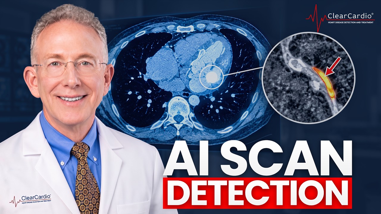
Dr. John Osborne walks through real-time cardiac CT scans of two patients, showing how AI now measures plaque volume to a tenth of a cubic millimeter. Soft plaque, which calcium scores miss, appears early in heart disease. The scan also catches incidental findings like fatty liver and osteoporosis.
Chapters
However, when plaque builds up and develops, the stuff that is laid down initially is invisible on a calcium score because it's lipid. We call it soft plaque, cholesterol plaque, lipid-rich plaque—all synonyms. That is invisible on a calcium score because it's a calcium score. As the plaque gets older, calcium will begin to develop in that those plaques. Then we can see it on calcium score. This is Dr. John Osborne, the preventive cardiologist that is the chief medical officer at Clear. Clear Cardio. I am really excited today. We have a fantastic program. We have some wonderful people joining us. They just got their scan with Clear Cardio, with our approach. If you have the disease, if you have plaque, if you have the most minuscule amount of plaque, we can both detect it, we can measure it, we can monitor it. But most importantly for you is we are really on the verge of helping to eliminate the disease. Plaque, atherosclerosis, that still kills today about 45% of us. And many times the first symptom of heart disease is death. Sit back, relax, and we're going to take you on a beautiful ride through the hearts of Floyd and Claire today. So there's your liver here, there's your spleen, stomach is here, and this is your left kidney. There's your left adrenal gland here. See a little bit of the pancreas right here. And we can also measure this. So, this gives you an idea if you have fatty liver, which is very, very common. So, that looks good. So, no issues there. Left kidney looks good. So, these cysts are just fluid-filled cysts. I'd say probably at least 10% of us have one, sometimes a lot of them. But we can actually— how we determine it's a cyst, we can look at the density of that, and the density says it's a cyst. So, this is the density number.
It's nothing you worry about, or you don't need to do the liver cleanses or the Yeah, it's fluid-filled cysts.
Probably, they saw it years ago, probably the same one. Hello everybody and welcome. This is Dr. John Osborne, the chief medical officer of Clear Cardio, and I have something really exciting I wanna share with you. We now have a newsletter. So this newsletter is all of the great science that I look at every day and read and scour the medical literature for important studies, important outcome studies, new tools, new drugs, new data on nutrition, all of the different things. that I commit my life and have committed my life to and passionate about in helping me to further preventive cardiology, from new diagnostic tools to new blood tests to new medications to the latest on intermittent fasting and dietary approaches to help people live longer and better. So it will all be in our newsletter. It is great reading. The price is right too because it's free. So please subscribe to our newsletter. Go to clearcardio.com or you can click down below and get access to our newsletter. Looks like the aorta is just a little On the larger side. Usually the a— this is the ascending aorta, the part of the aorta coming right out of the heart.
That means right to you, right?
It'll then— okay, exactly. The ascending aorta is the aorta, the big vessel coming out of the heart. That's about an inch and a half or so. It'll come up, it'll loop, give off the vessels to the head, neck, and arms, brain, and then loop and then head down to the belly. So this is the ascending aorta here, and then this is the descending aorta. But all that looks good. There's your aortic valve. So if you had leakage, narrowings, congenital abnormalities, malformations, or rheumatic deformity, we'd see all that. All that looks good. So let me tell you about mitral prolapse. So about 20 years ago, there was a paper published in the New England Journal of Medicine. So they looked in the prior 15 to 20 years, right? And so 35, 40 years ago, of all of the people having an echocardiogram, an ultrasound of the heart, we diagnosed mitral valve prolapse in 40%.
Wow.
4 out of 10.
Yeah.
Okay.
Wow. They then looked, so ultrasound got better, our definitions got better. They then looked in more contemporary echoes. Now it's about 1.5%.
Wow.
So 2 things may have occurred. I'll give you your choice. One, there's a massive epidemic of mitral prolapse and the vast majority of everybody got better, or else we way, way, way overcalled it. We could also look in the left atrial appendage. This is a little, I always think of it kind of as a lagoon, if you will, But that comes off the left atrium, and that's where blood clots like to form. So we can look at that, and that looks good. Particularly people who have atrial fibrillation, that irregular heart rhythm that occurs more often as we get more mature, those clots form in that little structure right here.
So no clots?
No clots. Wouldn't suspect it, but we can double-check on that. The left ventricle looks great. No old heart attacks, damage, enlargement. Right atrium, left atrium all look okay. No fluid around the heart, no pericarditis. So all of that looks good. No holes in the heart, congenital abnormalities.
How do you decide which image to look at? 1,600 images of the—
So this is you shown in 3 dimensions here. So there's your breastbone, there's your ribs. Now I will say, we'll get a little more quantitative in a moment, but as you look at the ribs, so there's your ribs right here, right? So the ribs are bone, right? So they're full of calcium, so they're pretty dense, right? And I will say, if you look at the bones here, you can see they almost look a little washed out. Yeah. See that? So that's an implication about the bone density. Yeah. Okay. In a moment, we'll do some quantitative stuff. But just looking at this, you can see, yeah, I wouldn't be surprised if he had some osteoporosis, right? Because normally the ribs, see how they look right there, pretty dense like a rib. And you can see towards the ends, they kind of get washed out.
Yeah. But, and they don't They don't test that on the test. They always test the hip bones and the back.
Right. So any bone can fracture.
Supposedly full-blown.
Yeah. But, you know, where it really makes a difference is the hips just because of the weight that they bear.
Yeah. That's where they test it.
There are 2 things, the hips and then the vertebra, right? Because you can also get compression fractures, which are really, really painful. So literally the bone just collapses, right? So you can certainly get rib fractures from osteoporosis. You can break any bone. But the ones that kind of really matter, we see most often involved with osteoporosis would be the vertebra and the hips.
Yeah.
But we had one guy that we diagnosed with osteoporosis who's a guy because it does affect men. He didn't know it. He'd never been tested for it. And he actually broke a rib without any trauma because—
How would that happen with no trauma?
Right. And what happened was he fractured his rib. The rib broke, and underneath each rib is There's a little, you can see kind of a, see this little indentation here? So in that indentation lies an artery, a vein, and a nerve. Okay? So every, on the bottom of every rib, there's an artery, vein, and nerve. So what happens is he broke the rib, which then disrupted the artery and the vein, and he ended up with a 4-liter effusion in the right lung.
Ooh.
And they said he had pneumonia, right? And then they sucked out 4 liters of fluid from his right lung. So he just thought he had pneumonia. Right. Well, he came in 6 weeks later to get a scan. We looked at his heart, obviously, but as we looked— as I was looking at this, I go, you broke your rib. He said, what? Not that I know of. But as it turns out, 6 weeks beforehand, before the pneumonia— and you can kind of tell the age of the fracture by how it looks. So you could say it wasn't an acute fracture, it wasn't an old fracture, so it was probably 6 weeks or so. Anyway, what happened was he broke his rib without any trauma.
Mm-hmm.
And had a lot of pain. And then he developed the pneumonia. Okay. So they never figured out he had osteoporosis and he fractured a rib until six weeks after later when we put in our machine.
Wow. Yeah. That was David Ledbetter, right?
That is correct. Yeah. Oh, his video.
He's the one that got me here indirectly because then he called David Frost.
Ah, got it. Got it. So there's your scapula there, breastbone. There's the descending aorta heading down to the belly. In the back there running next to the spine. There's the A sending over to here. And those are the big ticket items.
Yeah, okay.
But I will, I'll make some movies here along the way so I have some movies to show everybody here.
Doctor will be interested. She's real interested in the osteoporosis. There's some new technology now with that too.
Yeah, there's fortunately, the good news is there's a lot of very, very effective tools, medications to treat it. What I'm going to look at now is these are the slices we were looking before. Remember, this is 3-dimensional imaging. So here these slices are like this. Here, I'm going to take you and slice you front to back. So this is your diaphragm here, spleen, liver, right lung, left lung. We'll blow you up here. And there's your breastbone. Breastbone there. So there's your spinal column right there. There's your spinal cord. And then we can take this, make some measurements here. So this number here, 174. So if it's— now again, osteoporosis can be a little spotty, right?
Mm-hmm.
It could be worse in some areas and not as bad as the others. That 174, if it's below 100, that's 100% absolutely osteoporosis. If it's above 200, that's great. In the middle, but your 174, you can see the bones look fairly bright, which they should. So at least in the spine, I would say it's probably okay. But again, in the hips, could be a whole different story.
Yeah.
And then lastly, before we move on, there's your esophagus. We can do what I call the virtual endoscopy, when you take that video camera, put it down your stomach and esophagus. We can do the same thing for free. So this is just a poor man's endoscopy here. So if there's tumors, masses, cancers, varices, hiatal hernias, anything like that, we can see. And all that looks good.
Yeah, they were talking about doing one on her and I said, you wait a minute, maybe you won't have to get that done.
Yeah. So at least the esophagus looks fine. So, and then there's that little cyst I mentioned there. So why'd they want to do an endoscopy?
No, it wasn't that. It was the biopsy.
Oh, biopsy. All right. So we'll move on here. And I will say, so all the non-cardiac stuff, we have a radiologist look at that. I just take responsibility for the blood vessels. But doing 20,000 of these or so—
You get where you— yeah.
Yeah. I picked it up by osmosis. So I always have a gentle rivalry with my radiology colleagues to see if I know things that they didn't. All right. So we're now going to move. This is actually the low-resolution data. Now we're going to move into the high-resolution data, which is the heart. Okay? And her heart rate was absolutely perfect. And what we're going to do now—
Was that on the medicine that I had to take?
Yeah, the little cocktail that I gave you. Yeah, so we kind of— everybody gets a little different cocktail depending upon the heart rate, blood pressure. So I, I'd like to get the heart rate low 60s, 50s or so, and yours is 58. Gives us beautiful pictures. So what's going to happen now in the background is the computer is doing computing work. It's going to identify the heart here, and then you'll see in a minute It'll remove all the other structures around it. Identify all the blood vessels. There's your spinal cord there. We can remove that. Put it back. There's the liver here. These 2 vessels right here— oh, nope, disappeared. Okay. Anyway, never mind. So what we're looking at now is just the coronary arteries. So there's the aorta, that big vessel coming out of the heart, and then these are the coronary arteries. And we can, just for fun here, I will put the rest of the heart back just to see this. You can sort of see that. So there is your heart. And make a little movie here. So that is you.
All right.
So there's the left ventricle, right ventricle, right atrium, left atrium, aorta. And then these vessels right here are the ones we're going to look at with a microscope in a moment. Make a little movie there.
Yeah, I think the girls' biology class—
Exactly.
She can take it in.
So this is the right coronary artery that goes to the Right inside of the heart. We're going to take that vessel in here. That's the same vessel, but now we've laid that vessel out in a virtual plane, which you can't, but we do.
Okay.
And we've then taken that vessel and cut it right through the middle. So we can see not just the channel where the blood flows, the lumen, but also we can see the wall of the vessel because that's always where plaque starts. Okay?
Okay.
So that's what that would look like if we did all that. Please don't try this at home. And then over here, we've taken that vessel and we've straightened that vessel out. So this is the magic of computation these days. I call this cardiology CAD/CAM. Then we can take that vessel where these little green arrows are, and this will then create a cross-section of that blood vessel. So this is the cross-section right here, and then these are 1, 2, 3, 4 millimeters upstream and 1, 2, 3, 4 millimeters downstream. So we're going to put all this together, and here we're going to fly virtually right through your right coronary artery. coronary artery. So now this is where I have to make an apology. So in the old days, when I started doing this 25 years ago, this is, this is as far as we could take it. Yeah. About 4 years ago, we developed all these cool AI tools. And what the AI does is all of those millions of pixels coming out of the machine that make up this 3-dimensional high-resolution image, as it turns out, all of those millions of pixels encoded are encoded electronically with over 2,000 shades of gray. When we look at this, we call this grayscale, whether it's a CAT scan, an MRI, an X-ray, or whatever. Our eyes can see or delineate about 50 shades of gray. However, every pixel, technically voxel, little boxes, is encoded with over 2,000 shades of gray. So imagine we can see red, green, and blue. Imagine we can see ultraviolet, infrared, radar waves, gamma waves, gamma rays, microwaves, et cetera.
Right.
So we go from red, green, and blue to seeing the entire electromagnetic spectrum. The channel is completely normal until very late into this. So the vessel can— will grow outward to accommodate that plaque, to warehouse the plaque. Now, it can get to about double its size, okay, the entire vessel, right? Want clarity on your arterial risk? Click now the link in the description to schedule your Clear Cardio patient assessment call. We'll review your risk factors, see if our advanced program fits you, or point you in the right direction. So that's what AI does. So it literally can see things— that was my apology— That I can't see. Okay. Furthermore, if it does see anything, it will then quantify whatever it sees, the plaque, down to a 10th of a cubic millimeter, which is an almost infinitesimally small volume. And all of that was impossible to do up till about 4 years ago. So that's what the AI now will happen. So when we get the AI data back, it's gonna look at this. I'm gonna say this vessel here looks perfect. I don't see any plaque at all. However, I will tell you the AI is better than my eyes.
It's gonna come back. Okay.
So we'll see. Though I will say, having done hundreds of thousands of these AI exams, here's the data. Because plaque is so prevalent and so penetrant, you know, well, it is the leading cause of death in the US. It's the leading cause globally. 1% of us, when we use AI, will have a zero score.
Right.
1%. 99% of us have a non-zero score. Okay. And particularly if you, on the other extreme, we can now, because we have the ability to identify and quantitate it very, very accurately of the 99% of us that do have some plaque based on AI, that a third of us, because we now can, we have a staging system based on how much plaque you have, right?
Yeah.
So 0 is 0, and then we have 1, 2, and 3. So mild, moderate, severe, okay? Based on how much plaque we detect on AI. So a third of us, no symptoms, feel fine, normal stress test last week, I like to say, right? Which are not very sensitive at all. A third of us, when we do these AI exams, will have stage 3 plaque.
Okay.
A third of us. A third, right?
I just last month went to my cardiologist and had zero calcium.
On the calcium, right. So remember, calcium score is good, right? If you have a zero calcium score, your arterial age is 39.
Okay. And I doubt it, yeah.
However, when plaque builds up and develops, the stuff that is laid down initially Is invisible on a calcium score, right? Because it's lipid. We call it soft plaque, cholesterol plaque, lipid-rich plaque, all synonyms. That is invisible on a calcium score because it's a calcium score. As the plaque gets older, calcium will begin to develop in those plaques. Then we can see it on calcium score.
Okay.
And about 10 to 30% of us will have a zero calcium score, which is good. I'd rather take zero than not zero. However, 10 to 30% of us will have a zero Calcium score and yet still have detectable plaque. And as it turns out, the calcified plaque is, and I'll use an analogy I use all the time, that is the cinder cone. That is the extinct volcano. The lava that I really care about, the active plaque, the growing plaque, the plaque that is, that can rupture is the term we use, break off, leak off, pop like a pimple, whatever, that causes heart attacks is all the invisible lipid-rich plaque, soft plaque, right? Got it. Okay. So on a calcium score, I'm only seeing the tip of the iceberg. I'm only seeing the calcified plaque and I'm seeing the plaque that I really don't care about. No, if you have calcified plaque, 100%, how did it get there? We, it went through ladders.
Well, it had to go through the calcified plaque.
Exactly. So again, it's a helpful test. And the way I think of it is think of the calcium score like a mammogram. This is the biopsy, right? Because mammogram, we go, looks okay, looks not right. You get false negatives, false positives. When we do this, Yeah, I mean, it's binary, right? You got it, you don't have it. The rest is all—
You do a good job explaining it in layman's terms.
Just a little practice.
Yeah, when people stare at you like, what'd you say? You explain it well.
Well, I do like to say I am a pretty good translator between medical-ese to human.
Yes, not everyone.
Yeah, a lot of my colleagues just don't get that.
They don't translate. Yeah.
So, and I'm going to tell you, having done thousands of these, I have to tell you, every time we do it, I still remember the first time and it was just like, yeah, yeah, what? So that's why I call this the virtual autopsy. Yeah. Up until we had this data and then the AI added to it, the only way we could get this data would be an autopsy. Right. And we'll do that today.
Yeah.
So there's the vessel. And particularly when we look at the AI, you'll see The lumen, the channel looks beautiful, but plaque always starts out in the wall of the vessel. Okay. And early on, literally for years to decades, if plaque is getting deposited, the entire vessel will grow outward. So the channel is completely normal.
Right.
Until very late into this. So the vessel can, will grow outward to accommodate that plaque, to warehouse the plaque. Now it can get to about double its size. Okay. The entire vessel, right? Then the plaque begins to grow inward. We will not have symptoms, chest pain, angina, flunk a stress test, until we already have a 70% blockage.
So this is all on the inside?
Yeah. Right. Right. So, so when it causes a blockage, we get symptoms, chest pain, angina, flunk a stress test. But that does not occur until we already have a 70% blockage. So that's way, way late into the process. With this, we can see what's happening in the wall of the vessel. And even, you know, again, with a stress test, you gotta have a 70% blockage before it becomes abnormal. Plus when we do stress tests, there's about a 25 to 40% false positive rate where the stress test looks funky. Probably had friends.
Yeah.
Yeah. Right. I had a stress test. It was abnormal. They go to the cath lab and say it was fine. Right. Right. 70% of the time when we take people to the cath lab, we don't see any blockages. Right. 70% or 25, 40%, you get a false positive, which generates a $20,000 invasive heart catheterization with lethal complications.
Right.
25 to 40% of the time, even though there is a severe blockage, the stress test looks fine, which is actually even—
Yes. And you go home and fall out.
Exactly. Right. And the thing is the plaques that rupture, right, that cause the heart attacks are never 70%, almost never. They're 20s, 30s, 40s, 50s where there'd be no obstruction of flow. Your stress test would be fine. You have no symptoms. Those are the plaques, particularly ones that are more full of lipid or cholesterol that rupture. And that's how you go from, I feel fine, normal stress test last week, because I have to stick to my family history. And then you have a heart attack because one of those 40% plaques full of lipid, fragile, buttery lipid stuff will rupture. And within seconds to minutes when that plaque ruptures, your body responds with trying to form a blood clot. And that is the final common pathway into what causes heart attacks.
Heart attack.
Or if that— Or stroke.
Exactly. Same exact process, right? So, the same thing occurs in the blood vessels of the neck and we call that a stroke.
When we get to me, that's what I'm going to be very interested in, in my aortic valve. Did it have calcified and shut down the— Blood flow, the reason that they replaced it.
Yeah, well, we'll be able to see all of that.
Right.
And I'm sure—
I had to have some lava in there at some point.
And I'm sure before your aortic valve, right, you had a heart cath done, right? So the SOP in the old days was, even though we know you're going to the OR to replace the valve, we would do a heart cath to make sure you didn't have blockages, right?
The heart cath was at the last minute, wasn't it? Yeah, he didn't have symptoms. He had none. He was asymptomatic.
So, so what's interesting is, and I've been doing cardiac CT for 25 years, for my patients that have valve disease that need surgery, none of them go to the cath lab because we use this for 5% of the cost and none of the risk, right, to know if their blood vessels are okay. Yeah, right. So, uh, the, the next vessel we're looking at is the LAD. That vessel goes down the front part of the heart here.
Okay.
And again, take a look at that vessel here. So we'll have that here shortly. So that's the LAD. And then the other major vessel going to the left side of the heart is called the left circumflex vessel. And now sometimes we use 2 different AIs. There's the one based on this workstation here, and then there's a separate AI they use for that quantification. What happens, and you'll see that area here looks a little funky. Well, sometimes the computer gets confused. And if you look at this, you can see the vessel actually goes this way. So it got confused and it followed the wrong vessel. So that vessel is fine, but it looks a little funky because the AI, this AI, chose the wrong vessel to follow. And you can see it just makes a little curve there. But here, so there's actually the left circumflex. The AI got a little confused. And this AI is usually pretty good. It's very, very rare when it does this, but you can see it actually chose the wrong vessel. vessel to follow, but here we can kind of do it manually. And all that looks good. So I would say by eye, all the vessels here look great. Okay. We'll see what the AI says. Now remember, only 1% of this are completely zero when we use the AI. Okay. And then I'll show you just a couple other quick things here. We get this data for free. This will be particularly relevant to your husband. The computer will take the heart, chop the heart up into 17 pieces, and then measure the wall thicknesses throughout the entire heart. This is very helpful. There's a lot of conditions that can cause the heart to bulk up, to get thick. High blood pressure being by far the most common, and yours looks perfect, right?
Good. I've had blood pressure since I think I was in my early 30s.
Well, and obviously it must have been well controlled because, you know, if the blood pressure is not well controlled, like any muscle, when it has to work harder, it bulks up. And we can see that when we measure the heart, the thickness.
The heart muscle hardens and it doesn't pump properly.
Exactly, exactly. We can also then, from all this data, determine the exact size, dimensions of the atria and the ventricles, All of those look good too. So no enlargement of any of those chambers. And then we can also look at the valves. So here is what I call the surgeon's view of the aortic valve here. So the aortic valve is a tricuspid valve, meaning 3 leaflets. So there's 1, 2, 3. So it kind of looks like a 3-leaf clover or Mercedes-Benz sign, or if you're from the '60s, a peace sign. So whatever. So that's your aortic valve. So that looks perfect. So no narrowing, no calcification, no leakage. No congenital abnormalities, rheumatic deformity, etc. So all of that looks beautiful. And we can also look at that same valve in the X, Y, and Z axes. So there it is from that angle, and there it is from that angle. And we can also do the same thing with the mitral valve. So this is your mitral valve here. We can fly through it. So if you had congenital abnormalities, prolapse, narrowing, calcification, leakage, we can see all that. So here we are flying through it. So the nice, the cool thing with this is we get this incredible data that we could never get with any other tool. Looking at blood vessels in the heart, being able to see what's happening in the wall, that was impossible until we had CT. The AI will allow us to see stuff we couldn't see and then quantify it. But the other thing we get out of this is basically a very, very high-resolution echo, right? And we get that back through.
Yeah.
Yeah. So we can look at all the structural things, the valves, the sizes of the heart, all those things. With our echo, we get basically all of that data for free. And that looks beautiful. So no prolapse. I like that. Okay. Gotta keep you healthy. Take care of me. And you can see the mitral valve— this is diastole when the heart is filling— is open. So there's the anterior leaflet, the posterior leaflet, and then during diastole, the inward valve is closed. So there's all the little trabeculations, the papillary muscles, the chordae, all those little structures there. So looks great. Okay, so we'll see what the AI shows as far as that, but really looks pretty phenomenal here. Or as I love to say, you're just as beautiful on the inside as you are on the outside. Your husband told me to tell you that.
Thank you.
And I will make sure that we'll send you home with either a thumb drive or a disk with all of these pictures and the data and the lung pictures.
Yeah, one thing I found, because I had a disk that came from somewhere and I don't know if it's the different formats or what, But the doctor I went to, and it was after the pneumonia, they said, we can't read this disc.
Right. So what happens is all of this data is encoded in— so there's like JPEGs, right? So for visual pictures and all that stuff, all this data is encoded in a format called DICOM. So that is the universal language that all medical imaging is encoded in, in DICOM. So what you need is a DICOM reader that reads that data. Now, The disc that we create will have a DICOM reader on the disc.
Wonderful. Okay.
Right? However, anybody that has any DICOM reader can read any disc. There's lots of different DICOM readers, but the disc we give you will have the DICOM reader on it. But anybody like— so if like radiologists, all the data they get, no matter which machine took it, which CT, MRI, all that data comes to them as DICOM data, then they have I was surprised. So yeah.
And then my family doctor, when I gave her the CD and it was from the— she says, I don't even do a CD reader anymore. I'm like, okay. Yeah. So I don't know if some had gone to the thumb drive or what.
Yeah. I mean, we can do either.
And then the other, then the pulmonary doctor said, well, we can do it, but we can't read. And this was from an emergency center in town. They could not read that particular CD.
Well.
So anyway, I just—
Sometimes when they cut the disc, it'll have the DICOM data. Sometimes they don't put the DICOM reader on there. There's some generic DICOM readers.
Is it a problem to get both? To get the thumb drive and the disc?
No, you can do both.
I mean, if it's fast enough. But I was surprised because that was 2 doctors that could not read that disc.
So either it didn't come with that software on the disc, or maybe they didn't know to boot up that software and then use that to read the disc.
And this was a hospital.
Yeah, the radiologists, I mean, because they look at this and use this all the time, they always have their DICOM reader.
Well, who would have thought?
All right, so that is you. A couple cysts in the liver, no big deal. We looked at the bones, we looked at the esophagus, that little schmutz in the right lower lobe there, but the heart looks phenomenal. Looks beautiful. Don't miss this other episode right here on the screen where we will reveal the missing answers you need to know about true cardiovascular prevention. And if you're ready for absolute clarity on your arterial risk, click Click the link in the description to schedule your Cleer Cardio patient assessment. We'll look at your specific risk factors and see if our advanced program can help you.