The video provides a detailed clinical description of venous insufficiency ulcers, covering typical anatomical locations (medial lower leg, medial malleolus, anterior shin), ulcer morphology (superficial, irregular, moderate‑to‑high drainage), wound‑bed appearance (beefy red granulation, possible yellow fibrous coating), and the consequences of inadequate dressing absorption (edge maceration). It further outlines the spectrum of skin changes associated with chronic venous insufficiency, including cellulitis, dermatitis, scaling, varicosities, edema (often indurated), hemosiderin‑related hyperpigmentation, and lipodermatosclerosis (fibrotic, indurated skin). The second speaker shifts to professional development, urging clinicians to pursue wound‑care certification, highlighting its career benefits and the increasing accountability of Medicare and other payers for wound outcomes. The speaker promotes Woundeducators.com as a resource for certification and ongoing wound‑care education.
Chapters
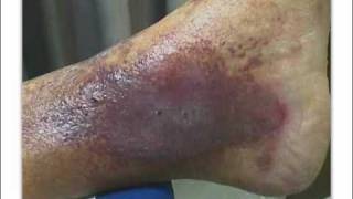
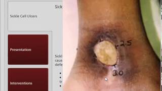
Venous insufficiency ulcers are usually located on the medial aspect of the lower leg or medial malleolus. It is also common to find venous ulcers in areas exposed to trauma, such as the anterior shin. Venous ulcerations do not occur on the plantar aspect of the foot and and rarely occur above the knee. Venous insufficiency ulcers are generally superficial, irregular in shape, and have moderate to high amounts of drainage. If the dressing does not absorb enough wound drainage, the wound edges will be white and fragile due to maceration. The wound bed contains beefy red granulation tissue, but may take on a ruddy appearance. A thin yellow fibrous coating may cover the wound bed, giving it a glossy look. Venous insufficiency is often associated with many skin changes. Cellulitis, dermatitis, and dry scaling skin, which may cause intense itching, are common. Superficial varicosities and evidence of previous ulceration may be present. Lower extremity edema is usually always present. In cases of longstanding venous insufficiency, The edema may become firm or indurated. Initially, skin changes may include a subtle erythema. With chronic venous insufficiency, the skin becomes stained and more darkly pigmented due to hemosiderin deposition. Hemosiderin is a byproduct of the breakdown of red blood cells, which have been forced into the interstitium by venous hypertension. The term lipodermatosclerosis is used to describe the hyperpigmentation and accompanying erythema, induration, and plaque-like structural changes that occur due to longstanding venous insufficiency. The skin and subcutaneous tissues are more fibrotic and less elastic than healthy tissue.
To learn more about moist wound healing and other key concepts in wound care, you may wish to consider becoming certified as a wound care specialist. The benefits of wound certification are immeasurable, both to your own career and to the standard of care that you can offer your patients. And because Medicare and other organizations are now holding healthcare professionals responsible for outcomes in wound care, there has never been a better time to become a wound care specialist. Woundeducators.com is dedicated to helping you take this all-important step in your career and to help you handle any type of wounds you may encounter in your daily practice.