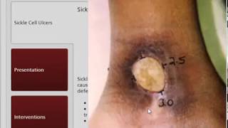
Pressure ulcers form when soft tissue compresses between bone and a firm surface. Spinal cord injury patients, hospitalized people, and long-term care residents face the highest risk. Prevention relies on positioning, mobility, nutrition, and risk assessment; wounds not healing in two weeks need reassessment and possibly electrical stimulation or surgery.
Chapters
Pressure ulcers are localized areas of tissue necrosis that develop when soft tissue is compressed between a firm surface and an underlying bony prominence. Patients with the greatest risk of developing a pressure ulcer are those with spinal cord injuries, hospitalized patients, and individuals in long-term care facilities. There are several risk factors for pressure ulcers: sustained pressure, shear and friction forces, moisture, impaired mobility, malnutrition, impaired sensation, advanced age, and a history of previous pressure ulcer. The majority of pressure ulcers can be prevented by using pressure ulcer risk assessment tools to identify at-risk individuals. Once identified, at-risk individuals will benefit from interdisciplinary interventions designed to prevent pressure ulcers, including education, positioning, mobility, nutrition, and management of incontinence. Pressure ulcers may be classified using the Integumentary Preferred Practice Patterns, the staging system, or both to better coordinate patient care among different disciplines. Pressure ulcer assessment instruments provide a consistent means to describe various aspects of pressure ulcers and identify changes in wound status. Keys to local wound care include protecting the surrounding tissue, addressing the wound bed, minimizing pressure and shear forces, and educating the patient and caregivers. High-tech support surface equipment is not a substitute for vigilance and holistic patient care. In addition to local wound care, physical therapy interventions for patients with pressure ulcers may involve therapeutic exercise and functional training. Pressure ulcers that do not show evidence of healing within 2 weeks should be reassessed for alternative or adjunctive interventions, such as electrical stimulation. Those patients not responding to conservative treatment options should be referred to a physician for further medical testing. Surgical interventions, including debridement or musculocutaneous flap closure, may be beneficial for patients with deep for extensive pressure ulcers.