The video provides a concise clinical overview of pyoderma gangrenosum (PG), a non‑infectious, progressive necrotizing skin disorder of unclear etiology. Approximately half of patients have an associated systemic inflammatory disease (ulcerative colitis, Crohn's disease, lupus, rheumatoid arthritis, etc.), while a quarter develop PG after trauma or surgery and another quarter have an insidious onset. PG most commonly affects middle‑aged individuals and is thought to involve neutrophil dysfunction. The classic presentation begins as a painful papule, vesicle, or pustule on the lower extremities or trunk that rapidly expands into full‑thickness ulcers with violaceous, undermined borders, purulent/hemorrhagic exudate, and rolled or indurated edges; systemic symptoms such as fever, malaise, or myalgia may accompany the lesions. Management combines systemic immunosuppression (primarily corticosteroids) with careful local wound care: gentle limited debridement, avoidance of aggressive surgical debridement, topical antimicrobials for secondary infection, and moisture‑retentive dressings. Adjunctive measures include limb elevation, gradient compression, negative‑pressure wound therapy, hyperbaric oxygen, and, after medical stabilization, skin grafting. Healing is slow, recurrences frequent, and pharmacologic therapy is typically continued for 6–12 months post‑closure.
Chapters
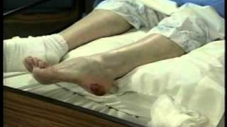
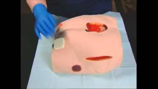
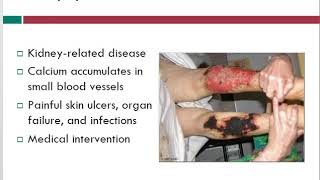
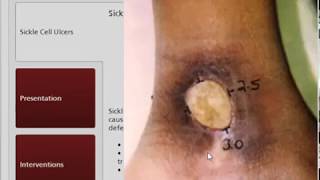
Pioderma gangrenosum is a noninfectious progressive necrotizing skin condition. The etiology of pioderma is unclear. Half of patients have a systemic inflammatory condition such as ulcerative colitis, Crohn's disease, irritable bowel syndrome, lupus, or rheumatoid arthritis. About a quarter of pioderma cases begin after some sort of trauma or surgery. But another quarter have an insidious onset. Pyoderma most frequently affects patients who are middle-aged. The pathology of pyodermic gangrenosum is poorly understood but is believed to be due to neutrophil dysfunction. The classic presentation of pyoderma gangrenosum begins as a small, very painful papule, vesicle, or pustule on the lower extremities and/or trunk. While a single lesion is possible, generally there is a cluster of ulcerations that rapidly expands. Pyoderma rapidly progresses to full thickness ulcerations with irregular violaceous borders which have purulent and hemorrhagic exudates. Undermining is common. The wound bed is eschar or slough covered and the wound edges may be rolled. The purry wound is indurated or boggy. The patient may also have fever, malaise, or myalgia. Pyoderma lesions are erythematous due to inflammation, but may also have a blue hue from epidermal necrosis. Immunosuppressive and anti-infective agents are commonly prescribed. Corticosteroids are used to control inflammation. Local wound care includes gentle and limited debridement of slough and necrotic tissue Extensive sharp or surgical debridement is contraindicated because it increases the inflammatory response, leading to severe exacerbations. Topical antimicrobials are indicated for sensitive bacterial infections. Gauze or moisture-retentive dressings should absorb wound exudate and protect the surrounding tissue. Extremity wounds may benefit from elevation and gradient compression. Negative pressure therapy and hyperbaric oxygen have been used with some success. Wounds that have stabilized with medication have been treated successfully with skin grafting. Wound healing is extremely slow. Because recurrence is common, physicians generally continue pharmacological management for 6 to 12 months after wound closure.