Epidermis Layers | Layers of Skin | Wound Care Education
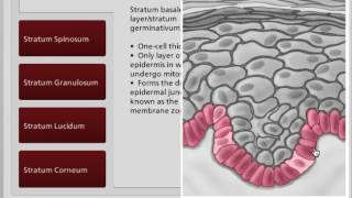
The video provides an instructional overview of the epidermis, describing its overall thickness (0.06–0.6 mm) and the five distinct layers that reflect stages of keratinocyte differentiation. It explains how new cells are generated in the deepest stratum basale, attached to the dermis via an acellular basement membrane that serves as scaffolding and a selective filter. The avascular epidermis receives nutrients by diffusion from the dermis. The narration proceeds through each layer—stratum basale, stratum spinosum (spiny keratinocytes), stratum granulosum (flattened cells with increasing keratin), stratum lucidum (thin, clear layer of dead cells found in thick skin of palms and soles), and the outermost stratum corneum (20‑30 dead keratinocyte layers that retain hydration and cause skin wrinkling in water). It also notes the variety of cell types present in the epidermis, emphasizing structural and functional aspects relevant to wound care education.