The video provides a comprehensive overview of hydrocolloid wound dressings for clinicians. Speaker A explains that hydrocolloids are occlusive or semi‑occlusive dressings made of gel‑forming polymers that absorb exudate slowly, forming a moist gel that promotes granulation, epithelialization, and autolysis. Advantages include thermal insulation, waterproofing, and lower infection rates compared with gauze, semipermeable films, sheet hydrogels, or foams. Various forms are described—thin, regular, bordered, padded, alginate‑combined, pastes, granules, powders, and specialty shapes such as sacrum‑shaped or beveled‑edge dressings. Indications cover pressure ulcers, minor burns, donor sites, venous insufficiency ulcers, and small cavity wounds, while contraindications include bleeding or heavily draining wounds, dry wounds, exposed tendons/fascia, infected wounds, immunosuppressed patients, and compromised skin integrity. Speaker B then details the practical steps for wound cleansing, selecting a dressing that extends 2–3 cm beyond the wound, applying the adhesive liner, leaving the dressing in place up to seven days, and removing it gently with or without an adhesive remover. The segment concludes with a brief promotion of wound‑care certification through WoundEducators.com, emphasizing career benefits and increasing accountability for outcomes under Medicare and other payers.
Chapters
Hydrocolloids are occlusive or semi-occlusive dressings consisting of a combination of gel-forming polymers that absorb exudates slowly by swelling into a gel-like mass. Hydrocolloids have different absorption capabilities depending on their thickness and composition. Upon removal, a residue commonly remains within the wound bed, which is often mistaken as a sign of infection. Although sometimes considered a low-tech option, hydrocolloids remain the most commonly used moist dressing in many settings for chronic wound management. Hydrocolloid dressings offer many advantages in wound care. When wound exudate is absorbed by a hydrocolloid, it develops a thick colloidal gel in the wound bed that increases the moist healing environment necessary for granulation, epithelialization, and autolysis. Hydrocolloids also provide thermal insulation and are impermeable to water, oxygen, and bacteria. In addition, hydrocolloids conform to body shape and provide a waterproof surface. Wounds dressed with hydrocolloids have lower infection rates than wounds covered with gauze, semipermeable films, sheet hydrogels, or semipermeable foams. There are a number of different types of hydrocolloid dressings, including thin, regular thickness, bordered, padded, or alginate combined. Hydrocolloids are also available as pastes, granules, and powder for use in cavities. Hydrocolloid dressings come in a variety of sizes and shapes, including the sacrum shape that is designed to fit the gluteal folds of the buttocks. Some hydrocolloids have beveled edges to reduce the tendency for the dressing to roll when placed in high-friction friction areas, and others have a smooth satin-like outer coating to decrease friction and shear. Hydrocolloids may be used for partial and full thickness wounds, both granular and necrotic. While primarily used to treat pressure ulcers, hydrocolloids are also indicated for minor burns, donor sites, and venous insufficiency ulcers. They may also be used in small cavity wounds as hydrocolloid paste, powder, or granules. There are a number of circumstances in which use of hydrocolloids is not appropriate. Hydrocolloids absorb fluid slowly, so they are not suitable for managing bleeding wounds or heavily draining wounds. Hydrocolloids should not be used on dry wounds, wounds with minimal drainage, or on wounds with exposed tendons or fascia. Because of their occlusive nature, hydrocolloids are contraindicated in infected wounds and should be used with caution on immunosuppressed immunosuppressed patients. Finally, because of the strong adhesive backings of most hydrocolloids, these dressings should only be used on patients with good skin integrity. To conclude, we'll take a look at how hydrocolloid dressings are applied in practice.
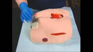
First, the wound should be cleansed with normal saline or wound cleanser and the surrounding skin gently dried, ensuring that it is free of any residue. Then the appropriate size and shape of dressing should be selected, remembering that the dressing should extend approximately 2 to 3 centimeters beyond the wound margin. Next, remove the dressing from the packaging and peel off one side of the liner. Place the exposed adhesive on the skin, removing the remaining liner while smoothing the dressing down onto the skin. Dressings may be left in place for up to 7 days depending upon the level of wound exudate. To remove a hydrocolloid dressing, press down on the skin outside of the dressing and carefully lift an edge of the dressing. Now gently loosen the remaining adherent edge from the skin by carefully working around the dressing. If necessary, an adhesive remover wipe can be used to facilitate gentle removal of the dressing. To learn more about moist wound healing and other key concepts in wound care, you may wish to consider becoming certified as a wound care specialist. The benefits of wound certification are immeasurable, both to your own career and to the standard of care that you can offer your patients. And because Medicare and other organizations are now holding healthcare professionals responsible for outcomes in wound care, there has never been a better time to become a wound care specialist. WoundEducators.com is dedicated to helping you take this all-important step in your career and to help you handle any type of wounds you may encounter in your daily practice.