A spoken YouTube case study (Gerald Bassleer) describing Case 10: Chaetodontoplus (marine angelfish) affected by head-and-lateral-line erosion (HLLE). The presenter documents external lesions on the head and lateral line, performed non-lethal scrapings and fecal examinations, and found heavy bacterial presence on lesions and intestinal flagellate parasites (Spironucleus, previously Hexamita, and Protopalena) in fresh feces at 100–400× magnification. He describes HLLE as a multifactorial syndrome (parasites, environmental stress, secondary bacteria/fungi) and recommends improving water quality, reducing crowding, and treating with antibacterial medication plus metronidazole; he explicitly mentions praziquantel? (no) — medication and dosing given: ferreltadone 1.5 g per 100 L and metronidazole, combined with probiotic/immunostimulant-supplemented food (Lapacho).
Chapters
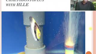
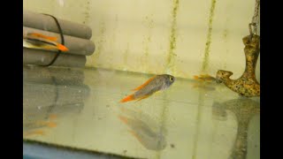
Hello, aquarium keeper or fish lover? Today, our fish disease. Case number 10, about the Marine fish Cetodontopoulos. Marines are suffering from head and lateral line erosion, a mix of infections, a serious problem in many of our varieties of marine fish. And you can see here on the picture that the lesions here on the lining, the lateral line of the fish, but also the head region of the fish. And the head region of the fish, that's also an extension of the lateral line. So the lateral line organ is here around the eyes, on the cheek, the gill covers, and here on the head. You can see it here on the picture. And you can see it here in the aquarium where we were called in to help the fish importer of this problem with several of this kind of fish. Here, the fish will commonly appear and be interested to eat something to show. And you can see here the lesions, all the leather line, all the erosions. It's a syndrome, it's an erosion. We will talk about what we find because we will examine this fish by taking some scrapings from the head and some excrements. We didn't sacrifice the fish. We didn't do an autopsy in detail. So we took a scraping of the fish from the holes or part of the area. If we can see if we could find something specific as parasites, what else? So the only thing we could see was a damaged area, but you could see no parasites, nothing making holes. The only thing we could see was a good amount of bacteria, which you can see here around the spikes of the scales. But that's all we could notice in our microscopic examination. So the next step was for us to examine the excrements. And here we collected fresh excrements on a plate. You can do it by squeezing the fish a little bit and squeezing it out. I didn't take a video from that because I was on location in Madrid doing that. So I was not having the material. But this we started to examine and look what we found. And in the microscope here, we could see at about 100 magnification, all those little buggers here moving around. So we say, well, let's go to a higher magnification of 200, 300. Nearly here we could see all little parasites. And these are Spironucleus, used to be called Hexametha. But here, all those little parasites were in between excrements found. And this is a typical way to find these parasites by taking some fresh excrements. But I must tell you, take fresh excrements, because if they're not fresh, you will not find the parasites or you find other parasites which are not the main pathogenic parasites. And here we saw something in between, something we will show on the next video. Well, we see here the parasites of Spirynucleus. And we see here one coming here and one coming here. So I was surprised to see something more, not one pericyte but also a second one which is called Protopalena. So we said, well, let's examine more details of other parts of the excrements. Because sometimes we found the spiral nucleus, the very tiny ones in specific parts of the intestine. I might explain that in other cases to come where I take a whole experiment and show you what we can find in different parts of the excrements. But usually here the spinal cord, spinal nucleus are sitting together at the same part of the intestine. And then we looked further in the excrements and we found these Protopalena parasites, the more elongated here, that's at 100 magnification. And see them moving around. They're very active, moving around. And you can imagine what this do in the microbiome and to the gut lining of the fish. I explained it in one of my first cases what head and lateral line erosion is as an impact. I think it was case number two or number three. Check it out. I had more details on the whole problem, what heterolateral line erosion is all about, about disrupting the immune system. Because the microbiome. And I explain you here later on about this case. Here, look, here is a parricide dividing, which is interesting to understand what has happened here. More magnification. 400, 300, 400 magnification. These are parasites moving around, very active, permanently eating, looking for food. They are like a big group sitting together. So they can easily be identified just by taking a scraping, taken a fresh sample of excrements. And here we show the one which is dividing. Because these parasites multiply very fast just by dividing. Here we see it happening while looking at the microscope. That's why they multiply so fast and can become a very nasty infection very rapidly within a few days and causing a lot of damage to the fish. At the damage of the fish is losing immunity and showing the heterolateral line erosion. And it's usually a combination. Like I explained in the previous case that there are parasites, there's some environmental factors playing a role. There are some bacteria or other viruses or fungi in the water. And altogether it's a combination that makes the disease as an outbreak and the fish show particular symptoms. So for the treatment of this co Infection. First of all, think about good water quality. Maybe do a good water change here. Or do a proper filter cleaning. At the same time is very well preparing the best conditions for your fish. Avoid crowded conditions. Avoid too many fish together, because then the disease can spread easily. Apply an antibacterial medication because here bacteria are causing the lesions you not a parasite. So you can use in this case ferreltadone at 1.5 grams per 100 liters. Use it in combination with the metronidazole. Because the metronidazole kills the parasites. The pericyte was inside intestine. You see off skinny. The fish already was. So the pericytes had a bad impact. So the metronidazole will kill the pericytes in the fish, but also in the water. Because they're spreading to possible other fish angelfish or tanks or other cytodontopolis. I also recommend to combine it with food. The food with pro and phytobiotics like we have in our Dr. Besley buys his food lapacho. With lapacho, it's for the repair and the fight of the parasites. The garlic is inside. And also we have immunostimulants to help the immunity of the fish. So the lapache would be a good combination together with the medications you give. And I can say avoid the spreading word Biosecure. Don't use the same net for this aquarium and for another aquarium, for example. It can be a risk of spreading. Similar symptoms appeared with a naso tank at the same location which was doing my work that day. And you will see when this fish turns around this nasal. We will see also that he started to suffer from heterolateral line erosion. Next to the lesions he had on the body. But you can see here the head regions and the head lateral line also starting to erode. And also he had already some damage on the body. So I might suspect that this had the same problem and that you can give it the same kind of medication. Because this disease, heteroderlan erosion is. Is common for tangs and angelfish and Chittatuntoplus and Zanclus, these kind of species. So that was this case. In the next case, number 11, I will talk about the Tropheus moliros. That's for the Chiclet lovers. More in detail coming soon. Thank you.