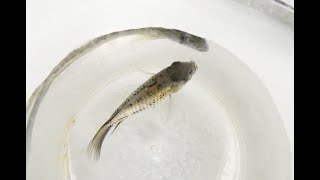
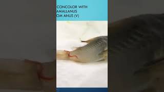
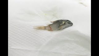
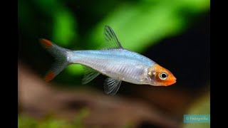
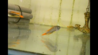
YouTube video (case #100) by Gerald Bassleer presenting a diagnostic case of Haludaria fasciata (lemon barb) suffering from multiple concurrent infections. The speaker shows clinical signs (skin wasting, emaciation, some healthy cohabitants), presents microscopic skin and gill scrapings identifying several parasites (Chilodonella-like ciliates, small flagellates, Oodinium/velvet) and secondary bacterial damage (columnaris-like bacteria, melanomacrophage centers in spleen and liver). He recommends removing severely affected fish, and treating with a combination of copper sulfate + formalin + salt (3 g/L salt), mentions commercial alternatives (Oodimore), and advises antibacterial therapy and supportive medicated food when needed.
Chapters
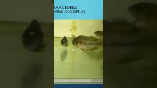
Hello aquarium lover. You're here today again and I hope you can celebrate with me my cases on fish diseases. Because today it is my 100th case of fish diseases. Yes, so far I reached the goal to have already 100 cases worked out for you. The aorist, the fish lover, who wants to learn more. Of course I will not stop because always I had to deal with fish diseases as a consultant trying to take care better care of you to give you the extra knowledge I can share with you with my experience in my work. So happy to do today my 100 case. And it's about the Halu Daria fasciata, which has an interesting mix of five question mark infections. The Hallucaria fasciata is the new name of the lemon bark. Here we see it swimming. You see how skinny that fish looks. Damaged, skinny, wasting away and dying. You see some other ones look pretty healthy and active, but every day a few were getting skinnier and they were dying and wasting away. Of course, that's the time you have to take action and to intervene and to sacrifice the badly suffering fish. To do a microscopic examination. And here you see a part of the skin scraping. You see all little creatures moving around in the water. Here we make the magnification a little bit bigger. You see there are some parasites moving around. You see the necrotic tissue with the bacteria. Some are Columnaris bacteria. You can see here at the edge, some Tubifex like growths, which is tubifex looking, it's the columnaris bacterial strains. But most important are of course these heavy moving around parasites. Well, let's take another scraping which we can see more easily those moving around parasites which are typical for a type of Hylodonella, which is a ciliated 1 cellular unicellular parasite which can locally be treated if you do it on time. There are some other parasites we could see also during that examination. And it's not always so clear to see that there's some small flagellates moving around. Then you have to go to a higher magnification like I show here. There's some small flagellates here, attached to to the necrotic tissue. But typical all those parasites here, the Hylodonella or the destroying the fish tissue, the skin, the fins and causing serious damage to the fish. And also during our observation we seen those dark brown parasites. Yes, they are not pigments. Some are looking like pigments, some that's the black spots here, that's pigments here, the black here, but here the brown ones. See that's These two brown ones here, I showed a higher magnification. Those are oodinium or velvet disease. And at the same time you see the Chylodinella parasites moving around. While oodinium is not moving around, it's penetrating and hooked inside the scale and of course causing local damage and the risk of bacterial infections. So you can see here the mix of infections. And when we examine the gills, we see all the black spots in here. Those are all oodinium. And you can see here already some damage which occurred to the gill tissue. And that is of course devastating for a fish. Like I explained in previous videos that that of actually the fish will die from. You see some air bubbles. No disease, no parasite. That's a typical air bubble. Well, the black spots are oodinium, non moving, attached parasites. Here are some loose because I took a tissue and some fall off in my slide here. See it at a larger magnification. You see here the oodinium attached in between the lamellae and attached in the gill tissue. At the same time we found melanomacrophage centers which developed due to bacterial infection. Here in the spleen, those are the darker areas. And here also the bleeding liver and melanomic. That means the macrophages are defending and trying to devour the bacterial infection and then making like clusters. And that's why those dark spots appear. Well, this is typical here in this case for a bacterial infection. It might also be other infections like a viral infection. But usually in our aquarium hobby, we find a lot of bacterial infection because of the tropical warm water that you can see in my previous presentations. Well, the Barb fasciata is the new name. It's the Barb. Well, what you can do is of course is remove the badly sick fish because they can continue spreading the disease when they are still around in the aquarium. And it's better to remove them because they can spread the disease and infect the other fish. And that's a risk. And you can do that by utilizing them with Narcomore Plus. First of all, I would start with to suggest. To make the suggestion to make a combination treatment of copper sulfate together with formalin and salt, 3 grams per liter. In my books I have a formula developed to show you how much copper sulfate to put in the formalin. Or ask your fish doctor, he might know it's available on the Internet and that helps you to control the oodinium, the velvet disease. But at the same time that formalin in salt and copper sulfate will control the other parasites like halodinella or if there is any costi or other flagellate parasites available. You also can try to use Oodimore, a medication from a quarterminster which also contains copper sulfate together with salt. Why first Oodynia? Well, because the gills are an important organ and this is usually the cause of the death when fish suffer from such as from parasite infections. When necessary, apply an antibacterial treatment if you see that the fish is not getting 100 cured. Well, we've seen it here on this fish with the liver and the spleen already badly damaged. And you can help with a fish food like the Dr. Basilisfood matrine that helps the fish to fight off the parasites and at the same time it repairs the fish and also the gut microbiome to develop a good immune system. Well, this I reached my goal already of 100 fish disease cases. So follow me on the Patreon. You will see all the cases there and more details on fish disease care. Well, thank you for watching. Thank you for being an attended aquarist. That helps to be on top of of fish quality control to become a happy hobbyist or a happy businessman when you're dealing with fish. Good luck at your fish care. Thank you for watching.