Gerald Bassleer examines Tropheus moliro with wasting and bloat caused by suspected Mycobacterium infection. Post-mortem shows damaged gills, fluid-filled bodies, and liver tubercles. Treatment involves oxytetracycline baths, probiotic feed, and strict disinfection to prevent spread.
Chapters
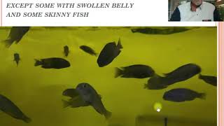
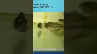
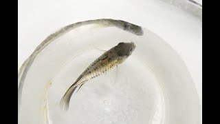
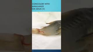
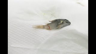
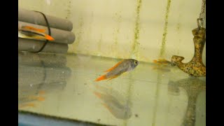
Dear aquarium fish keeper, now we will talk about fish disease. Case number 11 about the cichlid Tanganyika cichlid Tropheus bolero, which had a look, which is not very typical, but looking skinny and some had a swollen belly. Let me show you some pictures here. We could see if we took a photograph of the fish tank, that some of them had a little bit skinny here and here was skinny, but some other ones here but having some bloated, swollen belly. This one had swollen, bloated belly. This one looked a little bit skinny. If you look at the next picture here, we saw a bloated one. So here we had a bloated one. So we had a mix of, how can I say, of patterns of diseases. And the fish here you saw a few dying here very slowly, was not high mortality, but the fish were suffering. You could see it here. We see it on the video. The video is a little bit dark because of the lack of light. But you see some skinny ones here in the back. Fish which looks like they are wasting away. You see some, how can I say, some fish which are a little bit bloated, like this one here or this one here, and some looked skinny and wasting away. So it was not an obvious, clear symptom that we could notice on this fish. So what we did, we examined the skin, the gills, and the first thing we found on the gills, that some parts of the gills were destroyed. This is a kind of lesions we see on gills when fish suffer from internal problems. This is some small bacterial intrusion which can be an infiltration part for bacterial infection. But this is not a healthy gill. This gill looks more healthy here. This one has bleeding red here, some bleeding spots. So this gill is starting to get deteriorated here, another gill, we see that some parts are really badly destroyed by bacteria here completely wasting away. Also this gill started to be wasted away by bacterial fiction. If we examine the fish after dissection, we noticed that a lot of fluids escaped from the fish. The fish were having a fluid like a swollen belly with heavy fluid. That means the fluids couldn't get out of the fish, that some of the organs were not functioning well. So the fish was getting like a bloated swollen belly. Some of the organs look very pale, like we could see here inside of this dissected Tropheus moliro. When we checked the organs, we saw the spleen which had some reddish, inflamed inclusions. This is not a healthy looking spleen, can have different origins. And we checked the liver. We found very Typical tubercles. These are all tubercles, usually the tubercles. In most cases they are encapsulated by the reaction of the organ, in this case the liver, to protect themselves against infection. And in most cases, in studies from scientific reports, we found that about 80% of these kind of tubercles here are caused by Mycobacterium or the fish tuberculosis, Fortohetum or Marinum, for instance. Here we see another part of the liver, the Mycobacterium, look like tubercles, which of course were damaging the organs. So the organs were not functioning well. And some fish, they're getting skinny and some fish, they getting bloated. So these fish came from a breeder, a breeder in Europe. So what can you do about it when you have a problem like this? Well, first of all, apply an antibacterial medication. And we recommend in this case, oxytetracycline. Very high dosage, 3 to 5 grams for 1 liter. You can treat that during 36 hours and maybe repeat after one week. But not too much, not too long, because if you do too much, too long, you can create resistant bacteria. The very sick fish you should put to sleep gently. There are some products in the market that can put fish asleep. What you have to do is check your quality of the food you're supplying, because some fish food, like I explained in some of my causes of fish diseases, some of the fish food are carriers of mycobacteria. So avoid feeding live or frozen food for 30 days and try to work with a granulate food that has pro and phytobiotics that help the fish to defend themselves and to repair. For example, we have in this case our Dr. Basler biofish food, Fuco with fucoidan, which is a help in case of bacterial infections. Also, what you should do is avoid any contact with other fish with your nets or your hands. Make sure you also disinfect your hands, because when you have a wound, it can cause a lesion and can cause wounds infected by the bacteria, the Mycobacterium, which is a nasty bacteria. But if you work properly, your hair rarely get it or hardly never get it. So that was about the Tropheus bolero. In the next case I will talk about the Malawi Chiclet, a case of an alunakara, which has some patches, some damaged eye. So stay tuned for case number 12. Thanks for your attention.