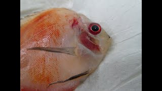
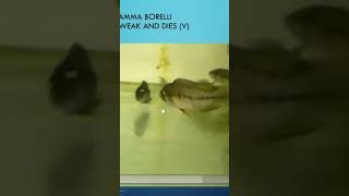
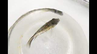
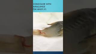
Gerald Bassleer examines tank-raised discus with wasting, pale damaged gills, and bloody organs over weeks. Microscopy showed dark filamentous infiltrates in gill tissue that may indicate a Branchiomyces-like fungal infection. Treatment options include formalin, malachite green, methylene blue, or antibiotics, though the breeder source may be the root cause.
Chapters
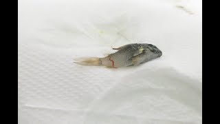
Dear Aquarius, dear fish keeper, and especially for those which are discus lovers, this is a beautiful T shirt with some wild discus. But today I will show you something else about discus fish from a breeder which was suffering from mortalities losses. In a couple of weeks and months we had a continuous incidence of fish which were wasting away and dying. And well, we did an examination and we made some conclusions, but not specifically exactly we could conclude that this was one disease or a mix of diseases. But anyway, I will show you my findings and maybe you have some suggestions or maybe you did some research yourself. And you can fill me in by sending a reply to this video I share. So I'll show you my findings in my PowerPoint that I prepared for you. It's my case 113 and it's about 10 crazed discus fish which had very bad gills and bloody organs. A difficult case. Why it difficult to define the exact path? Again you need specific lab research in some cases. But here we see that the tank raised pigeon bloods in a fish tank. Some become darker, they waste away and eventually they become very skinny and they die. You see them here, you see them here getting very skinny and not eating well. The bigger ones will eat more and take away more food. So there's a competition so that the weaker ones will finally suffer more. And the same was here with a malborder red species. See here this one is getting very skinny also. You see the respiration is very rapid and well they get skinny and they die. And it's one every two, three days, maybe every week. It's very regular. It's a very slow wasting away disease. So we, we took the fish and we examined and the first thing we found when we opened or removed the gill cover were pale gills. Well, normally the gills have a nice red color, but here you see some patches inside on the edges here. Well that, that's the first sign that you see. Well, this fish is suffering from some problems in the gills. And then we put it on the microscope. You see that the gills are very poor colored here. Here they're darker and more red, but here they are pale and not well formed. So we have to go more in detail and look at it. Here you see there is some excessive mucus on, on top of that and there is some big darker areas and some bloody hemorrhagic lesions into the gills. So the gills are in very poor shape. So we had to go in more detail and check that. We found some inclusions, some bad gills with add some specs inside. Very strange. I Could not say exactly what that was on this kind of image. But the gills is not normal. They should be normally like in between here, red like here, but not all those spots and specks inside. And then we go in another examination more larger magnification at 400. We soon do is darker hair things sitting inside in between. And in the gill lamellae. Is this a kind of fungus? Well, let's go in detail and see more parts of the gills. See, some of the gills are completely well infiltrated by a kind of organism. And it might be a kind of fungus. It's not a parasite. It doesn't move. It has not the features of a parasite. So we went in more research and I get you some more video to show. Here, look at this part here. It's all infiltrated with some of that organisms. And I always have a suspicion that it is a kind of gill of fungus which is not very common. Here we see it again here. I hope you can see it also here. I don't know if anyone has seen this one before because it was very unusual to find this. But I found it in a couple of different discus from the same breeder. Here we see it again. All those organisms, all here. A kind of fungus? Yes, maybe. Look at the gills here. Again my suspicion was to define it like Branchiomyces. And why did I think it was Branchi mice? Because I start doing my literature study, pull my books out of the shelf and look what studies have been published on this kind of incidents. And then I found a study which had also those gills look like here, all those black specks inside. It was a study done on also other cichlids, tilapia from aquaculture farm. So I will give the references on the study later on in my discussion on the treatment. Also the organs. You look at the liver here by the page of blood. And the spleen. They had all those melanomic centers, bloody red specks inside here the liver also the Marlboro red also in the organs, in the gut, in the intestine, ready bleeding maybe. Is that a virus or the blood spots baby virus causing that. But look, the whole combination. That fish is suffering from a very strange problem which I couldn't define with my naked eye. So that's why I had to go. Well, first of all, study on the gills. And it's branchia mesis, like here. You can try to follow that link here. Let me open it for you. Then you will see how it appears. And this is the branchimesis, like infection in a culture. Tilapia. And this is the study discussing that branchimesis like infection which was discussed in Israel in the laboratory. And well, you can follow the full text. And here is that picture I got for you in detail. So. And here are those kind of spores, parenchymasis spores, which we tried might have seen in our microscope for the discus. So that's. Now let's go back to my presentation here. Okay, that's not what I want to share. Well, let's go back to my PowerPoint. There we go. I have this. Share it again. I'm sorry. That's what happens when you start pulling around in your bike in my computer. Anyway, let's go to the details of that treatment here, like I said. Well, I showed you the link. Well, lab can help to identify the pathogenic agent. Well, you have to tell. Please check the gills, the internal organs. It suggests test here. A treatment with formalin malachite green, methylene blue, which is a good antifungus. Treatment for this kind might be a reason to apply together with Farnamore, which is also a possibility. Well, not together. FMC or Farnamor. Farnamor is a anti white spot in treatment that might kill. That kill fungus combined with veromorph, which is a kind of bacterial control. So applying antibiotic treatment might also help. Those are suggestions. Well, let me know what you think, what you find out. Well, it's complicated. Discus are very valuable and you must think about it. Well, feed the fish with our biofish food professional treat, which is a general help when fish are sick. And that has different functional ingredients in that treat. It's a herbal treatment and it's a garlic treatment, an aloe vera treatment and give that food for several weeks and no other food. And that might help you. The problem might be the breeder since both fish came from the same source. But the freighters stopped his business in 2022. Maybe because of the problems, I don't know. Also the energy crisis, the high cost of having a discus farm. It's not so easy in Europe. Okay, well, this is that picture again here of the branchiomyces. Well, get my books on fish diseases. I discuss other kind of fish disease. This one was very unusual, which is not published in these in my books because it's very uncommon to have found this kind of disease. But the most common diseases are discussed in my books available in different languages for you, you can get it from my website and follow my patreon to become a specialist if you want to get more training. So thank you for watching. It was a detailed study on. When I give you some other links, you can look that well, make up your conclusions and let me know your findings. Always interesting to discuss other opportunities. Thank you for watching.