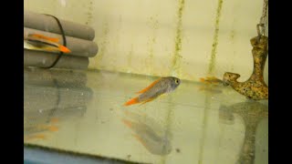
Gerald Bassleer presents Case #12: an Aulonocara (Malawi cichlid) in poor condition with multiple co-occurring problems. External signs included emaciation, skin patches, frayed fins and a cloudy/damaged eye; microscopy of a skin scrape (50× then 200×) revealed a moving flatworm (monogenean-like) attached to scales. Necropsy showed reddened, slimy gills, hemorrhagic intestine, fatty degeneration and liver lesions, and kidney melanomacrophage centers—findings consistent with bacterial infection and secondary parasitism. Recommended actions: systemic antibacterial treatment, antiparasitic for skin flukes, change to sterile granulate feed, and use of probiotics to support recovery.
Questions this video answers
50× and 200×.
Ferltadone 1.5 grams per 100 L or oxytetracycline 3 grams per 100 L.
Parasucantel 250 milligrams per 100 L.
Dr. Basler Bifishwood Fico.
Presence of melanomacrophage centers (MMC), indicative of prior bacterial exposure.
Chapters
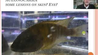
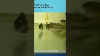
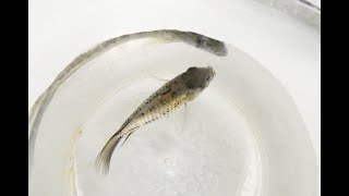
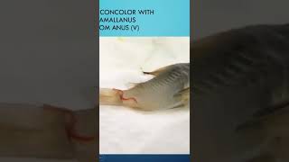
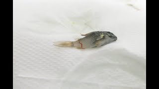
Dear fish keeper, today I will talk about fish disease. Case number 12, about the Olanakara. It's a Malawi shiglit and the Malawi shiklet, which I will show here today. He looks stressed, he is skinny, has some lesions, a cloudy eye, but no typical pathologic symptom. What we can see with the naked eye. So I show you some photographs and some of my microscopic observations that can help you. We saw the fish. It was obvious that a few patches on the skin could have been noticed on this ola narcara. And the eye seems to be somewhere damaged. Could have been a lesion of aggression or other cause. We could not determine, but we know that the fish was acting not so healthy. He you will see it, how the fish is swimming, how skinny he looks. You see some patches on the side. You see some of his fins are a little bit, how can I say, frayed. You see here a few patches here on the body. The fish particularly was very skinny. Well, I took the time for this fish to sacrifice because he had a lot of friends left in the tank. About 50 of his male and female friends were still alive. And what we found here is on the skin scraping here. It's at the magnification of 50. We saw a kind of Apache area here which was not clear for me. So we magnified it and we start seeing that it was a moving object. This was an organism. Very difficult to see at this magnification what it was. But it's not a white spot. It's not a small unicellular parasite like Caladonella or Kostia. So we go to a higher magnification of 200 and then you could see here it's like a worm type flatworm. A flatworm which sitting on the scales of the fish. Here we see it again, another type of coloration. You see the two pigment eyes of the worm, see the body shape of the worm. It's a typical kind of flat worm, a kind of monogenetic traumatode. Don't know exactly what species this. I'm sorry, I don't spend time and money to identify exactly what's the scientific name of this kind. But what I know is it's a flatworm living on the skin of a fish which is a kind of monogenetic trimatode that can cause damage. So what you want to do is to treat the indications we have. But we didn't stop searching, finding this worm on the skin. We also looked at the gills and we saw that the gills are pretty red and look Badly reddish, stressed gills, a lot of slime, some bacterial intrusion here. So this. So the healthy gills were not there. So the fish were also suffering from a problem in the gills. And this could have been caused by a problem by the inner organs. If we look inside in the intestine, we found bloody patches in the intestine. This is in hemorrhagic lesions. Could have been caused by bacteria, for example. It's like diarrhea in a fish can be causing this kind of symptom. So the fish might have had food which was in bad condition. Or the fish was in polluted water with too many bacteria. But this is not normal to have these bloody streaks in the intestine. If we looked further, we saw a lot of greasy fatty degeneration. Also the organs here, like the liver, had red patches, holes and had fatty degeneration. Could have been caused by a bacteria. So the organs were in poor shape. And what was very bad. When we checked the kidneys of the fish. In the kidneys we found all those kind of MMCs, melanomacrophage centers, which is a reaction on bacterial infections in most cases. Most cases bacterial infections. So this is going to be due to a history of the fish already had been imposed to exposed to bacteria or exposed to food with bacteria microbes into the food. So that could have been his suffering. And this was making the fish very weak. When these kidneys like this look like they fail and the gills will fail because the gills will have to take over the job of the kidneys and then being stressed. So kidneys are bad, the gills are bad. Fatty degeneration. And then on top of it, they had some skin flukes. So this is a complex problem for this kind of fish. The first thing you should do, apply an antibacterial medication like ferltadone, 1.5 grams for 100 liter or oxytidrecycline, 3 grams per 100L. Check for sure your quality of food frozen or fresh. And try to avoid feeding frozen or fresh food fly food. During the next 30 days. Try to give granulate foods which are free of microbes. Meanwhile, try to kill the parasitic flatworm on the skin because that's also hurting the fish and causing lesions. You can use Parasucantel at 250 milligrams for 100 liter. I try to help your fish by feeding it with pro and phytobiotics to help their defense system and for repair here. In this case, I would recommend the Dr. Basler Bifishwood Fico so this complex problem which we found on the Olonacara might give you some idea what you can do about it. And maybe you will encounter also this problem in the future. The next case, number 13. I will talk about a marine fish. This Abudevduf Syanaus, the blue damsel. An interesting case. See you next time. Bye bye.