A case video by Gerald Bassleer (with son Baron Basler) showing diagnosis and treatment of disease in Botia morleti. Presents clinical signs (fish lying on bottom, red/bleeding patches), performs a rapid field dissection and wet-mount microscopy of gills, liver, spleen and intestine, identifies gill rot and heavy bacterial presence, and recommends antibiotic treatment (and supportive measures) with reported good results.
Chapters
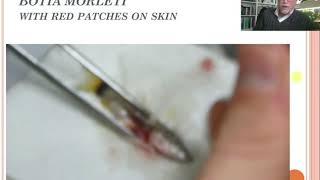
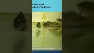
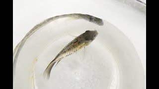
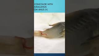
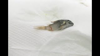
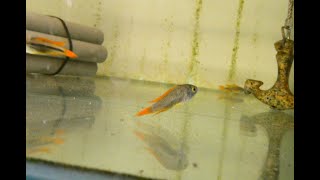
Fish disease, case number 14, about the boccia morleti or the skunk or mouse boccia, also called where the fish is laying on the bottom, red skinny patches and dying. You see here the red skinny patches on this Boccia moraletti. And here we see the fish in the aquarium. Many of them look okay, but you see some are a little bit nervous and stressed, but some are laying here at the bottom, not moving. Here is also one here up front and showing some red patches. So some were dying, but in general, the group of fish were still okay. So that means we could still treat on time if we act properly and do a proper examination of the few sick fish. Because the group of fish might all be infected or at least 10, 20% today and maybe in 2, 3 days there might be 40, 50% being infected with the disease, which we gonna now try to check for. We found that we took out the fish. We also saw some red bloody markings here at the anal part and at the belly part. So if we see this kind of red patches on the skin, we might certainly expect a bacterial infection, might also be parasites. But anyway, our macroscopic observation will help us. And now today I will show you my son, Baron Basler, who will show you here the dissection of this Boccia marletti. We show it in a very simple way. This is a very rapid diagnosis by taking skin scrapings and we taking out organs. And I show you how simple it can be done. Because my son is 25 years old, he's a young biologist, and he's getting the training to develop the tools of doing a proper diagnosis in a very simple but fast way. Because time is money. It's important that you spend not too much time in the facility on doing very technical research on Finch diseases. What we need to know is on a simple, fast way to do a correct diagnosis. And that means taking out the organs which we want to examine or can examine with the help of a microscope here. He's taken out the gills, he's taken out the gill tissues just to make sure that he has enough material so he can check for the possible diseases or pathology on the organs. And it takes altogether a few minutes. So these are the guilt tissues. Now he's going to do opening up the fish by doing a dissection with his scissor, a very simple way where he starts up front and he's cutting carefully so he is not damaging the internal organs. So now he will cut off or try to cut away the flap of the skin tissue and the muscular tissue. So he has a free view of the organs which we want to examine. Because examination of a fish totally is the skin, the gills and the internal organs. The organs internally is usually the intestine, the spleen and the liver. And with those five examinations, skin, gill, liver, spleen and intestine, we can find most of the problems. So he's taken out here the different organs. Now he's taken away here the intestine and also the spleen is attached to it. You can see the redness there. And I think once he took already a part of the sample of the liver. Now he's going to take another part of the liver which is there in front of the fish. So this is simple. We don't use any coloring techniques. We just use raw pathology. We call it field dissection, field microscopy. And anyone can do that. It's just an experience for you to try it out. Now he's adding some drops of water. Usually we use the water of the aquarium because the water of the aquarium has the same type of condition that the pathogens are used to live in. Now he's putting up the small coverslips. We prefer to use small cover slips on the different organs because it's easier to do a different examination of the organs. So he put it all on one slide. So it took him about three minutes. So he's now squeezing it a little bit because we prefer that the organs and the tissues are flattened so they are much easier to observe in the microscope. I will show you the pictures of the microscope. Now. What have we seen on this fish after Bernd Basler has done his examination and dissection of the fish? Well, we see that the gills, the reddish bleeding parts. We see here a gill which is rotten away and damaged. Here also is a part damaged, more gill tissues. You see here a badly damaged part. Here is. That part is damaged here. This tissue is gone. This tissue is partially gone. And usually it's. We can see more in detail that it's usually caused by bacterial infections. We call that gill rot. Also, the organs on this fish were bloody organs. We saw the bloody spots on the fish outside. Well, the organs were bleeding. Here you see a very heavy spleen bleeding with some red markings inside. Also, the liver has very red markings. So we might suspect bacterial. It could be viral infections. But we have no tools to examine this. It costs too much money, too much time. So in this case, usually we can only treat bacterial infection. That's why we try to look for antibiotics. And usually the bacterial infection comes Secondary after the viral infection. And a viral infection we cannot control. We only can control the secondary problems and we can strengthen the fish to fight off the viral infection and to become immune. We also find a lot of bacteria, internal organs here next to the liver we found a lot of rod shaped bacteria and round shaped bacteria, so different kinds of bacteria, which is probably the cause of the infection. That's why we decide we're going to have to treat this bacterial infection with an antibiotic. Of course, first of all you have to check the origin of the problem. Maybe it's the water quality, maybe it's the food source. A bad food like frozen or live food with polluted with too much microbes, poor water conditions. Maybe the fish has been shipped in a lot of stress with a lot of ammonia, which can cause these bacterial infections. So we recommend to apply here an antibacterial medication like oxytocin or androfloxacine or other antibiotic and feeding the fish with pro and phytobiotics to help the defense system of the fish and to help prepare and we use for this case the Dr. Basler, Baishvur, Fukkel. So this was a case with dissection examination of of this boat shell Moraletti with red wounds, probably most likely caused by a bacterial infection. So we treated with antibiotics with good success. We had good luck with that because we anticipated very soon in the beginning of the infection. The next thing we will discuss next time will be an infection of a discus albino discus. So see you next time. Thank you for listening and watching.