Gerald Bassleer examines an albino Discus fish from a distributor showing emaciation, eroded scales, and gill damage. Necropsy revealed enlarged kidneys with calcium deposits and dark lesions in the liver and spleen indicating systemic infection. Bassleer suspects contaminated food and recommends antibacterial treatment, dietary changes, and probiotics to support recovery.
Questions this video answers
Case number 15.
May 18, 2021.
"oxytocin, cyclin or other antibiotic" (as stated by speaker).
Melanomacrophage centers (MMCs).
Chapters
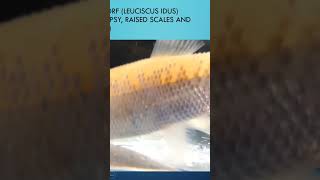
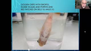
Today we discuss fish disease. Case number 15. Yes, it's already 1514 you might have seen, and more to come. Well, today it's about Discus Albino. I received from a large distributor of discus fish. And the fish was getting skinny, emaciated, and few fish were dying in the tank. Although the fish looked very nice and big. You can see it here, and you see here the skinny abdomen, how badly the fish was getting and that his health was somewhere affected. Some fins were a little bit damaged, but not serious. No fin rot we could see. We inspected the fish in the lateral line and there seems to be some little erosion, which you can clearly see here on this lateral line and a little bit here also. But there was not very much else we could see on the fish. We took some scales and we examined the scales and we saw some scales which were damaged, eroded. See, there were some holes here, the sides were like eroded, you can call that erodent scales, damaged scales. Here we see another scale, you see the large erosion here. And there is sometimes a weakness of the fish in the formation of his scales, and it is due to an internal problem. We find that back in other cases and which I will show you in the near future, when the fish is suffering from organs who are not functioning well. And it seems to be a weakness of the fish to get his minerals and calcium, etc. Up to the scale formation. So the scales look damaged, showing an internal problem. Also the gills look like a deformation or disforming. You see, the gills are not nicely firmed here. They seem to be clumped together. So there seems to be some damaged gills. So the microscope tells us more. You can see here the bloody appearance of the gills are bleeding. And here we see some gills which are eaten away or completely eroded. Damaged bacterial infections were occurring. You can see this one here also. So the gills were having a serious problem. So the fish was also suffering by heavy respiration. And when we opened up the fish after the dissection, we noticed that the intestine was full of food, very dark. We saw very large kidney and we saw the empty abdomen. So the fish had some organs that were not well formed. And so we examined under the microscope and the first thing we see that the intestine was very well, how can I say, completely filled with food, that it was not digested. We could find that back under the microscope and you check the excrements. We found poorly digested food when we took some of the organs out here, we took some of the samples. Here we see at the left the spleen, this is the liver. At the right there is the kidney. So these three organs, you see already the dark, dark spots, and that is not normal, those dark nodules or tubercles, what else we can call it is not normal to have this in the organs. See, what we see here in the liver is those dark spots is melanomicrofarg centers, MMCs, which is usually, in most cases a bacterial cause, sometimes a viral cause. But, well, I tell you, it's difficult to find virus back in our fish. We have so little tools to examine. Here we see a bad spleen. It's the same problem with the MMCs, the Milano microfox, etc. And we also see here the clocking up of the veins. In the kidney you see some calcium formation. So the kidneys were not working well. So the organs were very badly affected. So treatment for this kind of problem. Well, the first thing we have to ask ourselves, how did the problem start? Was it stress? Was it the food causing it? The water conditions? Maybe. But we suspect in this case the origin to be the food. A food with a high microbial pollution, which can come from frozen or live food. We recommend in this case to apply an antibacterial medication like oxytocin, cyclin or other antibiotic and change of course, the food source. We recommend to feed the fish with pro and phytobiotics to help the defense system to repair the fish. And Dr. Basley Baijer food has therefore the forte. The grapefruit seed extract, the moringa, the herbal or the foucault. So what is the best? It's hard to say, you know, it's already difficult to get the Discus to eat dry, granulate food. But you will eat eventually, when he is hungry enough and still want to live for a longer time. The fish which are too weak, well, that might be difficult to save. But think about it. Looking at the origin, it's the microbial pollution which usually comes from a kind of food, frozen or live food, which is not well prepared, not well cooked or not well kept. So this was my suggestion for this problem of this Discus albino. The next case will be about the giant Betta giant Beta female. You see picture here, that's the next case, number 16. Well, see you next time. Stay tuned. Bye bye.