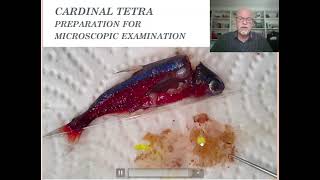
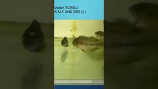
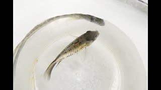
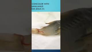
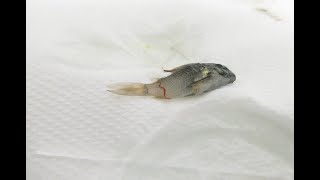
YouTube video by Gerald Bassleer (Fish disease case #18) presenting a case of swollen, tank-raised cardinal tetras. The speaker documents external signs (large bellies, no raised scales), dissects small tetras to examine gills and internal organs, and finds widespread tubercles in gills, liver, spleen and gonads consistent with mycobacterial infection (fish tuberculosis). He discusses likely origins (vertical transmission, contaminated live/frozen food—mosquito larvae), diagnostic limits, zoonotic risk, euthanasia for severely affected fish, biosecurity, and possible antibiotics and probiotics.
Chapters
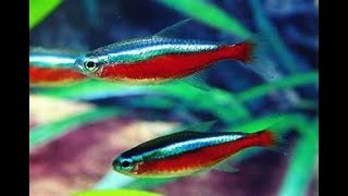
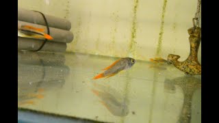
Fish disease case number 18. Today it will be about a cardinal tetra, a tank raised one. The fish is swollen, has a large belly and a few fish were dying. So you can see here on the. On the photograph. You can see here the big body of this carnotetra. Here is another one here, the two we have here. But they have no drops here, nor they have raised scales. So it's not like ascites or dropsy. It has another type of disease. Here we see in the. In the picture. In the video you see also some pop high. You can see this one. Here you see the big bellies for the wounds we selected out of the fish tank. And when we checked some of the tissues for the organs, we found in the gills that they were in very bad shape. You see some of the organs were here broken apart by most likely bacteria. And here also. So the fish was suffering from a bad gill. Tissues also we found, in some parts of the gills we found tubercles. And this is of course worrying. When you found tubercles in the gills, you suspect some more problems internal in the organs of this cardinal tetra. So we will show now a preparation how we take out the organs of the cardinal tetra and then prepare them on a slide. So it's like a study for you how you can do an examination on a fish which is 1-2 cm small. It takes about 1 minute. And explain you what we do when we dissect such a fish like that. The gills really have been taken. The fish is decapitated. You see, the head is off. And we use very tiny small scissors for this. And we start opening it from the lateral line where we have decapitated the tetra. And we take a very careful cut of the side of the body. And then we just open the tissue. We just pull open the body of this small cardinal tetra. Here we see it on a larger magnification. And we just use fine pencils for this. And then we see inside the organs. Here we see the swim bladder. Usually the swim bladder we rarely examine. There's rarely any disease we can find there. And we just take out, scrape out the tissue, all the tissue, the organs. See more there. That's the liver, which came out as last part. And that all we. I just collected on a glass plate here, a microscope slide. See the different tissues. Here is the intestine with the gonads. Here is mostly the liver on this part, in this we will put some water on. Usually I put water on from the aquarium, which is the same temperature where the fish has been kept in. And then we just put another slide on it, a glass slide. And we just squeeze it like they do here. We just squeeze it. And you can really see that the organs here, the liver, all has black, dark brown spots, also here and here also to the spleen also. So as you can see, this doesn't look normal when you see this kind of organ with all those brown dark specks. Well, this is what we can see in a microscope. We see here that liver which has those brown spots and those are all tubercles, plenty of tubercles in that liver. Looks very unhealthy. Also, the spleen has already lost its red color and it is loaded with tubercles. And it's also between the eggs, the gonads we found here also tubercles. So the fish was completely loaded with tuberculos in all the organs of this fish. Well, the fish was already a couple of years old, probably might be. And well, for a treatment for this kind of fish, which is already so swallowed, that's too late. You have to put the fish asleep because it's most likely tuberculosis. Of course, like I told you before in my previous presentations and lately I had a few on tuberculosis, but because it's a common problem, I explained it in my in my videos in YouTube. And mycobacteria is something that develops easily in our tropical aquarium and a laboratory can confirm. But like I tell you, who is going to pay €150 or €225 to find it out? Of course, a breeder should do that. Well, again, like I explained in previous explanations, the origin is usually the mother fish who passes it on through the gonads. And also live or frozen food with microbial pollution. And mostly it's from mosquito larvae. Mosquito larvae are very common to carry mycobacterium with them. And it cannot be killed by freezing. You only can kill it by, well, cooking the food. Euthanize the sick fish, apply biosecure measures, because be careful, it can infect other fish, but also can infect wounds on your hand. So you can have a zoonotic infection on your hands and can have some problems which you don't like to have on wounds you have on your body and particularly on your hands. You can try to use medications like erythromycin, tetracycline and minocycline and feed the fish with pro and phytobiotics. That helps in the defense system and the repair. I recommend our buy fish food Fuko for this, which is a big help to fish to survive better this problem. Well, like I said, I have a specific video on my YouTube channel about fish tuberculosis. Try to watch it and you can maybe learn something more about this nasty disease. The next video will be about swordtails. Look. Swordtails that have wounds. Okay, see you next time.