Veterinary aquarist Gerald Bassleer presents Case #20, a YouTube lecture describing an emergent physical disease in aquarium fish characterized by skin "vesicles" or bubble-like lesions. He identifies the agent as Dermocystidium (a mesomycetozoan-like organism), shows macroscopic photos and microscopic stages (plasmodia and spores), contrasts it with other causes (gas-bubble disease, Pleistophora, nematodes), and gives pragmatic guidance: quarantine/euthanasia of heavily affected fish, antifungal baths (e.g., Dysamore) to reduce spore spread, and prevention by checking sources and improving husbandry.
Chapters
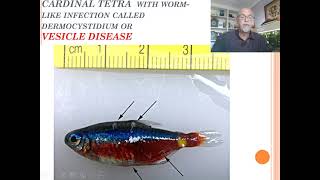
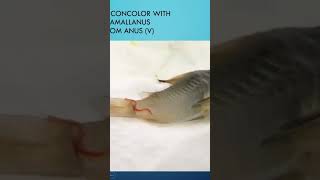
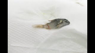
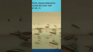
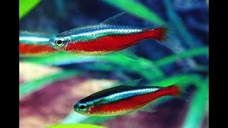
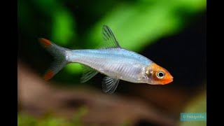
Hello. Today I will discuss with you my fish disease, case number 20, about the physical disease. I give it another name because it's an unusual disease we are dealing with in the last coming years, where the aquarium fish get large bubbles on their body, on their skin. We call it vesicles and some people say it looks like worms. So I will discuss several photographs and several fish I received during my work as a fish doctor. And then you got these kind of tetras, like a rummy nose or neon tetra. They have bubbles, kind of bubbles or physicals on their body. Or this rubinos. You see the different symptoms. I received this from a friend. This kind of bubbles here on the side, very strange appearances which we are not used to see. And it's something just recent. In the last couple of years we are seeing this more and more. And some people say, well, I see a worm, infectious. Look, they have worms here, there is a worm there, those whitish worms. But I tell you there are no worms there. Hear the same on this cardinal tetra. You see this worm like appearance, something sitting in a bubble, sticking on the gills or the fin or usually on the skin of the fish here on this black emperor tetra, a worm like infection. And I call it a vesicle disease to give it a name, because I used to give names when I give training to the people. I don't call it bubble disease or something like that because it looks kind of like a gas bubble. It's a physical disease. It's like a worm here. Yes, of course it looks like it. But a microscopic examination can tell us what's going on. We see the corydoras with many vesicles. Look at this, all on the body. And later on those white appearances, white worm like appearances of our features will come into the bubbles here on this Loricari day, all those bubbles appearing. It's not gas bubble disease. No, it's something else. And it's dermocystidium, that's the, how can I say, the parasitic infection. We call it sitting in a vesicle. Look at this here. We see it magnified. I received this from a colleague veterinary researcher in Brazil. And you see them here sitting all here on those bubbles. You see this worm like feature sitting in there. And it's a dermocystium. And I'll explain what dermocysteum is. See here again, really looks like needy, like little worms. Yes, that's why people try to use anti worm medications, but it's not working, unfortunately. You will do nothing with that. So this is what we see when we take a microscope, a macroscopic view. It's a kind of fungus or a sporozoa. There is still discussion among the scientists how do we catalog this kind of infection. It belongs to the mesomycetozoa. It's something like a mix between the fungus and the sporozoa. Here we see a larger magnification, the different spores. And what we see here in that bubble, usually it's the reproductive stage. Those are white hiphae. The white hiphae. This is what here looks dark, of course, in the microscopic examination. And that we called a plasmodium. And that contains many spores. And that's the way it will re infect other fish in the aquarium. That's how it will spread. This will break open and the spores will distribute and reinfect the other fish in the aquarium. Here we see another Dermocystidium, or physical disease in a pangasius. And this is the real gas bubble disease. Some plants appearing. This is on a marine fish so easy to diagnose. No. I can imagine that people sometimes have a wrong diagnostic observation that they call it like a bubble disease. But its microscope helps us. This also the white appearances on the skin. This is a Pleistophora sporozoa infection. I will explain that in one of my next cases. And here it's a white spot. It's called caused by Beta cercaria worm larvae. It's all something else. And here, this is a real worm. This is a real bubble with a real worm. When we opened that bubble, we found this large nematode, longer than the fish itself. And the rummy nose. This is coming from a wild rummy nose from Colombia. This is also a real worm infection. We open it, it can look like Dermocystidium or the physical disease. And we open it and we found a real nematode inside in this wild cardinal. Well, this is sometimes common. Well, not common. It's usual. We can find this in wild fish. Well, what can we do about a treatment for this dermocystein or physical disease? Treat the diseased fish in a quarantine tank, try to remove it and put it separate. Because in the aquarium, this diseased fish can continue to spread this fungus or spores. What else to the other fish in the aquarium? You also can treat the aquarium with an antifungal medication like Dysamore. We had some positive results to kill the spores which could be distributed. Follow the directions of the medication. If it will treat the fish which has already the big bubble or the vesicles with the large hifa, the large plasmodia. No, no, because it's there. But you can try to prevent the spreading. That's what you should do. Or the reinfection, the very sick fish you better euthanize. And then you also go to check where you get the fish from. Do you get it from a specific shop, a specific supply? Because usually the source is from the breeder. You can also cause the problem. But usually the breeder is breeding this disease in his fish tanks or in his ponds by having a serious problem, a poor condition. With that there's dermalcystium in the tank. Sometimes fish food can be the source of the problem. Several studies pointed out already many years ago. For instance, like Schipper Claus published in his book that Crustacea or Daphnia, like copepods, can be a risk factor for introducing this fungal or parasaic infection. You can help the fish with immunostimulants and recovery food, like our Dr. Baslior Baishi's food, Aloe vera or our professional treat. These help the fish to repair, to recover. But like I said, a very sick fish, very difficult. Try to separate the ones with the very large vesicles with the white look like worm infections separated and the carotene attack. And maybe it cures, it goes away and after a time it disappears. And then you can put the fish back after you might have been treated that with an antifungal medication. So it's something coming up. Try to follow your fish. Observe your fish regularly. Usually it appears between the same kind of group of fish, the same species, because it's from the same origin. Usually the fish come from usually Tardocardinal Tetrasomer Farm or the Rummy nose. And well, try to prevent the spreading. I hope you can be stayed informed with more news from my Patreon and you can follow me on my Patreon channel. Gerald Basler. I hope I can give you the best training for fish disease diagnosis. Learn more about fish diseases. Also my books, which you can order and the links will be presented below in my YouTube channel. So I hope I can keep you happy as acrist. Good luck. Bye bye.