Gerald Bassleer examines cardinal tetras with white patches caused by two infections: Pleistophora (neon disease) and Mycobacterium (fish tuberculosis). Mycobacterial infection is hard to treat, so management focuses on isolating affected fish, supporting survivors with probiotics, and considering antibiotics like minocycline, though results are limited.
Questions this video answers
Case number 21.
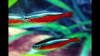
Cardinal tetra (Paracheirodon axelrodi).
Pleistophora (microsporidian spores).
Tubercles consistent with Mycobacterium (fish tuberculosis).
Dr. Basler Biofish Food Fuko, fed for 20 days only.
Chapters
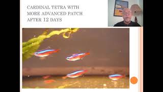
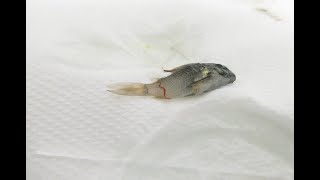
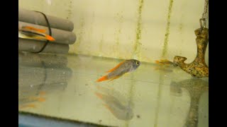
Today we will talk about our fish disease. Case number 21. Yes, already 21 cases. And shortly more to come. It was about a case of cardinal tetras, which were tank raised by an importer. And they were dying after a few days, 25 days with white patches. So I took some of those fish to my, my laboratory because I wanted to investigate what was going on during the weeks when the fish developed this kind of symptom. This is how it looks like. Only one or two in the batch of cardinals with this kind of patch of the body, the loss of the blue color. So something which seemed to be very common from certain suppliers, from certain regions, certain countries, I will not name a few. It's not so common from the wild. It's particularly tank raised cardinals, which seems to show this problem. And during the few weeks I had it in my laboratory, we did some testing on diseases. And afterwards we also did some testing on how can we treat, how can we make the fish healthier and try to survive this disease. Well, in the beginning stage, when we received the fish, see we had here one swimming here with that discoloration, that patch in the body, which looks very healthy, still swimming well, eating well. Look, all the others looking in good stage here. After day one, that's how it looked like. And after day 12, that same cardinal had a much larger patch which I show here, much more loss of the blue color. So the stages were advancing. So after 12 days, that's how badly it looked like. And the fish started to become more nervous, showing some stressed behavior, more separating from the group. Fourteen days again, few days later, the patch really started to grow bigger and larger and the fish were suffering more than the days before. And Even after day 23, here is 14, which I just showed here. At 23, it was getting more, larger. Nearly half the body, the colors were disappeared and a big patch was appearing. So that's day 23. So this is 21 days, a few days before I show here how the disease developed. You see the. If you can see the respiration of the fish very fast here, here see how he's suffering, also respiration. So somewhere the body was in very badly stressed situation. So that's the fish we're going to examine. And I took autopsy from the skin tissue of the tissue which we saw it was damaged with lesion. And we found all those little groups of spores, sporozoa, which is called Pleistophora or could be determined as Pleistophora, that's the neon disease. We found that in the muscular tissue and here we see them and high magnification 400. You see inside are many, many little spores, little parasites. In causing those white discoloration by the tissue of the muscular tissue. On the other hand you also found some tubercles also in the muscular tissue. This is a part of the spine. See here is the sporozoa, here is sporozoa, but here are tubercles. It's another kind of lesion usually caused by Mycobacterium. Here we see again the Pleistophora. And here we see some development here of a tubercle starting. So there's a difference in pathological observation. Here you see the spine of the fish. And you see here the sporozoa. And here we see some starting of tubercles. So this is a mixed of combination. We found here again another picture with tubercles and the sporozoa. The tubercles here, the darker ones and the more brighter ones are the sporozoa. So the fish was very suffering from two problems. And particularly the mycobacterium is a worrying thing because this is also killing the fish. While the fish with sporozoa infection can live a long time with that neon disease. We used to call that neon disease the white patches. But tuberculosis is something that the fish will waste away eventually because it's usually caused by mycobacterium or fish tuberculosis, which can be defined by a laboratory but very difficult to treat. On the other hand, also the parasitic infection is very difficult to treat. So what can we do with a fish like this? Well, the first thing you should do is try to discover as soon as possible this problem and put the fish in a carotene tank because they are spreaders, they can spread it to the other fish, particularly when the other fish are eating each other, particularly when they're eating the diseased or the dead fish. So remove it. So the badly damaged fish, when it's badly, badly damaged, you should euthanize it. You can use Narcomore plus from MyCorrh Munster which is a very tranquil, safe and animal friendly utenization problem. Solvation so that the fish is not suffering when you kill it. Treating while giving food with pro and prebiotics to restore immunity, particularly the remaining healthy fish. And we have a food which is called Dr. Basler Biofish Food Fuko where you can treat 20 days only with that food, no other, which helps you that the fish become more resistant to parasitic and bacterial infections. Particularly it helps the fish to recover during bacterial infections. Antibiotics well, best could be shown In a laboratory with an antibiogram, some people use minocycline or oxytecycline. It's a good choice, but to be honest, it will not so always perfectly control the tuberculosis because it's inside capsules for that bacteria cannot be reached with antibiotic and due to sporozoa infection, cannot be treated. This is a problem by the breeder, so the breeder should solve it. He should eliminate the origin of the sporozoa and select these breeding pairs to avoid that sporozoi spreads in the community. So this is how the remaining fish looked like after the 20 days of feeding, we had no more recurrence. We had here one fish with some darker patch. Here I show it again here. There was one with some darker patch, but he seemed to do well and he did not develop later on here. This one here, he did not develop later on any infection. And I had these fish done in my home aquarium for the next year. So it seems to be no problem. This is the same kind of sporozoa infection showed here in a Ruminostetra, a tank raised Ruminostetra. You can see this is typical neon disease caused by the sporozoa Pleistophora. Sometimes it's confusing to identify Pleistophora, so I always recommend use a microscope because here you see a white patch which is caused by Tetrahymena parasites. Or this is Columnaris disease, which is a bacterial infection, not a Porozoi infection. And this can be treated with a proper antibiotic treatment. So I explain more in my, in my training courses on fish diseases, how to treat columnaris disease. So a microscope helps you, like I showed you previously, to determine this problem. So I hope this case helped you to be well informed about the fish disease and how to solve this kind of problem. So thank you for the attention and I hope you stay tuned for the next cases to come.