This is a spoken-case YouTube transcript (case #25) led by Gerald Bassleer about a bacterial infection in Iriatherina werneri (a rainbowfish). Reported clinical signs: some fish become skinny, lose color or darken, show erratic swimming and sometimes flared gills; group behavior changes and mortalities occurred. Pathology: pale (decolorized) spleen with melanomacrophage centers, poorly functioning kidney with multiple calcium deposits. Microscopy: wet-mount of organ fluid at ~400× magnification revealed numerous rod-shaped bacteria and other transparent/ mixed bacterial morphologies. Likely cause: bacterial infection exacerbated by poor maintenance, overcrowding, contaminated food or stress. Recommended actions: improve water quality and feeding, consider antibacterial therapy (oxytetracycline), and supportive products (Dr. Basleer BiFish Fuko food, grapefruit seed extract, or professional treatments).
Chapters
Hello. Today we discuss our fish disease. Case number 25.
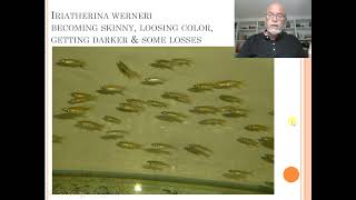
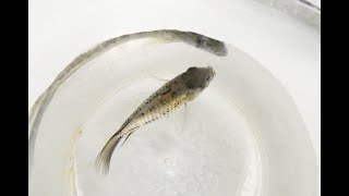
It's about the Erythrina vernery with a bacterial infection. We found the Eryterina veterinary, that some of them become skinny, they were losing color, some were getting darker and we.
Had a few losses.
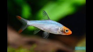
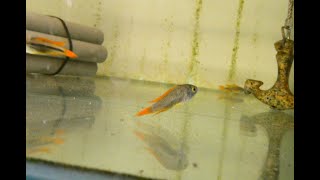
This group of Erythorinia, which we show here in the picture here below we.
See this darker colored one. Sometimes the gills are flared open, but not always. Sometimes erratic swimming here. And here's another darker one. And some look pretty healthy, very nice color. But it starts always with a view to show this particular sign, this particular symptom.
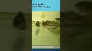
Here we see it again in the video.
Here we see some of the darker ones appear here below the skinny one, the dark one, you can see also the behavior is not that normal. Look like this one here, it's not so well, swimming in the group.
Well, these are the first signs you.
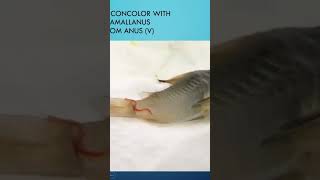
Can detect when there is a problem occurring in your fish, particularly when it's a group swimming, behaving fish, or when it's not behaving normally in the group. Well, this is a particular sign. We did some examination of the organs and the particular thing we found in the organs that here, the red part, which is the spleen, just without coloration. We take a sample, we press the organs between two glass plates and we.
Saw those melanomacrophage centers.
This is the kind of reaction most.
Likely due to bacterial infection.
You also see here the kidney of this fish with many calcium deposits, which is looking like a very bad functioning organ. The kidney is not looking well, the spleen is not looking well.
And when we examined the fluid between.
The organs, we saw many rod shaped bacteria here. We can see them all sitting here. This is about 400 magnification.
If you go higher magnification, you will.
Not see more than this.
So this is a bacteria we found. Also next to the rod shape, we.
Also found transparent spots. Different mix of bacteria we observed in this fish.
So our conclusion was that we found a bacterial infection most likely. Of course we could send it to.
The laboratory, but in many cases we don't do that. We look at the pathological sites, we avoid the time loss, or sending to.
The lab takes a few days and meanwhile the fish could die.
The origin of such a problem with.
Bacterial infection could be poor maintenance, overcrowding, polluted food that contains too much microbes, bacteria, a weakened immune system due to different stress factors.
Well, the first thing we improve the.
Conditions of the fish, improve the quality of the food, make sure you have.
Good water, you can treat with an antibacterial drug. Like in this case, we recommended oxytetracycline. It's still working in some cases. So it's an old drug, but it's still working.
And at the same time, try to.
Feed the fish with our Dr. Basler Bifish food Fuko. Or the grapefruit seed extract or the professional treat. So those are some recommendations for this kind of problem in this group of Ireturina veneri.
So more cases to come. Please stay tuned and I will show you more.