YouTube video (case #37) by Gerald Bassleer describing abnormal external growths on Chaetostoma (suction-mouth catfish) specimens reportedly from Colombia. The speaker presents visual observations, light-microscopy findings suggesting large enlarged cells with inclusions, and interprets the cause as likely viral (secondary tissue proliferation after stress). Recommended management focuses on improving husbandry, reducing stress, treating secondary infections, and supporting immunity via specialized feeds.
Chapters
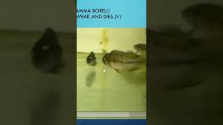
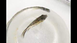
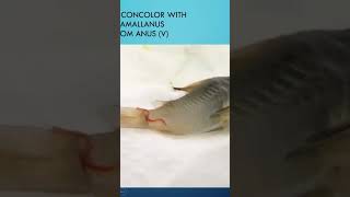
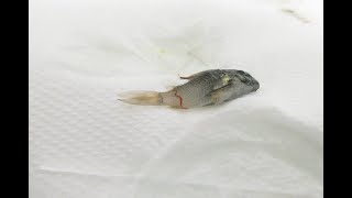
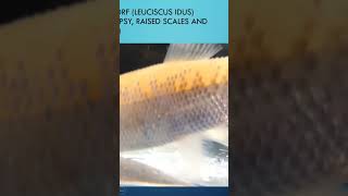
Hello, Accordion fish keeper Fish disease, case number 37 already. I hope you've seen the other 36 which I made because they might help you to understand fish diseases, how to diagnose, how to find parasites or bacterial infections, and what is the best ways to treat. So this is number 37 and this might help you to understand another specific case which I found on schetostoma, Schettostoma tomasi from Colombia, which was reported to me and given to me with some growth on the skin, some extra skin developments, growths outside on the body of the fish. And the first case, very difficult to see something. But you see here the little specks on this one here you see the specks here and here you see it here. Not easy to see. But if you observe well your fish, you can see that was something abnormal. It didn't look like white spots. It looked like some slime, some extra mucus, some extra tissue, some extra growth on the side of the fins. On the body of this cytostoma, you can see something on the mouth which is. I show you here more clearly. See here is some extra growth on the mouth, which is not normal on the head regions. And that was. The Akaris showed me this problem when he saw disappearing on the nose of the fish here you see this extra mucus growth hanging on it. Is it a parasite? Is it a something else? Well, that's why we use a microscope. And we saw the microscope, we saw this large tissue, like a big cell that has been growing, you see, like a lining here around. And in many cases I found in my, in my work as a fish doctor that this was caused by a viral infection. Virus can cause it. Well, we cannot see the virus as we know, viruses are very small. We learn that through the COVID 19 cases. You need a very good quality electron microscope to detect the virus. You can detect the virus with a good laboratory method, not by seeing signs and symptoms. This is a secondary problem. The primary problem is to make a correct identification. Here we can see the pathological issue of a very large cell which is enlarged by something inside. And in our experience in our fish industry, in aquaculture, in the hobby, it's in many cases caused by a virus. And usually this happens after stress, after fish have been put in bad conditions, bad handling, poor water quality, shipping handling, many things can happen, do this. Why the fish getting weak. And there's a possibility of infiltration of viruses in the tissue. Here we see also some strains growing. Tissue can develop to larger cells. We see some small little. How can I Say inclusions inside, which is hard to identify if you don't have an electronic microscope. And here we can see also kind of growth here. So we see some abnormal appearances in the mucus of the fish, which is not normal. And even on the hair on the fin, you see the growth Here you can see the whole tissue. Here. That's the extra growth, which is like cells which are infiltrated with virus, which are multiplying and causing the. Here you can see the inclusions of the directions of the tissue, sometimes a defense system. Here we can see some inclusions. And that can most cases in our experience caused by a virus. We found some studies on that, we explained the dead viruses. But I tell you, in aquaculture, viral examinations are not well funded, not well researched. So it's a suspicion that it's a virus here also here we see another kind of inclusion, enlarging of the cell, the genetic material that's sometimes multiplying and making extra growth tissue on the fins of the fish. So here is from another case, an historical example I had in my fish tanks a couple of years ago, which you see a very advanced case of those cell growths on a tail of an ancestress. This is a very large case. And it was the same kind of problem, because same kind of origin from Colombia in this case. So what do we do in the treatment with ice treatment for this kind of problem? Well, first of all, improve the living conditions, improve water quality, improve filtration, good water changes, avoiding stress, because usually it happens after the fish went through a lot of stress. And even if you work on these three things, you could make good water, good hiding places, pieces of wood, for instance, for the Schitt stoma, some darker water, brown water, not too much light, good food, everything. It might disappear by itself. Because to be honest, there is no treatment to kill a virus. You can treat the secondary infections. We learned that in COVID 19. We cannot kill the virus. We can help that the fish or the animal or the human being can kill, can protect themselves against the viral spreading or viral replications, and particularly helping the secondary infections with medications. And you can help to improve immunity, which is something I found very important in my work. When you give the immunity of the fish a good strength, a good strong immunity, the fish can fight off many diseases and can recover very fast. We recommend in our for the hobbyist to apply our specific Dr. Basler biofish food. Forte, which is with beta glucans for the immune stimuli, or the herbal, which has some four herbs inside to help fight off bacterial parasitic effect helps the fish to recover. Or professional treat. For the professional fish keepers, importers, exporters, pet shops, which is a mix of ingredients, broad spectrum treatment and use at least this kind of food for 20 days, no other food. So the fish have enough chance to absorb the essential micronutrients and ingredients to prove its immunity and to improve recovery. So this was another case I presented you. In the background, I see a beautiful skyline in the evening in Manaus, on the Rio Negro and Rio Amazonas. Enjoying one the past one of my trips in the Amazonas, which we learn where we learn about fish, about fish diseases, what they need. How can we help our fish to prevent diseases and to cure. I hope my information helped you today to become a good fish hobbyist. Thank you.