Study nr. 6 How do fish get Fish Tuberculosis or bacterial infection Mycobacterium
Gerald Bassleer breaks down how Mycobacterium chelonae infects bettas through different routes. Injections kill fish fast; oral exposure and skin wounds cause slow, chronic disease with granulomas. Poor water quality, contaminated food, and handling injuries drive outbreaks in tanks.
Chapters
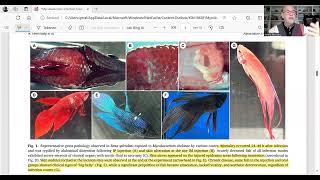
Hello, Dear aquarist, dear fish keeper, dear fish health manager, today I will present you a study that I found very interesting about mycobacterium, or fish tuberculosis in betta fish. Well, it's not only concerning betta fish that this discussion I will give you on this study is important to understand how can fish become infected with mycobacterium. And the study checked the different roads, routes or possibilities of fish infections, how they are introduced to the fish. So I will give you the most important details of that study which I find interesting in my kind of work. So I will share with this study here in my screen and I indicated the most important parts that might be interesting for us to understand. So it was about the consequences of an infection and studying the histopathological, pathological consequences in the Siamese fighting fish due to the exposure to pathogenic Mycobacterium via different routes. Well, the different routes is important to understand how our fish can become infected with mycobacterium. And as I indicated here in yellow in the abstract, mycobacteriosis is a chronic progressive disease in fish caused by Mycobacterium species. And it's typically characterized by granules, lomatous lesions, this cranulomas, tubercles, you can say, that illustrate its harmful pathogenesis, as I already give many different cases on fish diseases with these kind of cranulomatose lesions. Well, this is a study that elaborates more than that and maybe gives more insight in my findings as a fish doctor. And this study was initiated to understand the progression of mycobacteriosis in Siamese fighting fish or the betaspledins through the various transmission modes. Here in the study, the bacteria were administered by five different routes and the mycobacteria they collected from an infected fish. So they cultivated the bacteria and they administered that to different routes at three different dosages. And they used injections, intraperitoneal and intramuscular injections. They administered the bacteria to oral administration through immersion and also immersion with fish that had skin trauma. And they already could conclude that acute infectious, typified by short survival and non specific growth size and severe AGRA necrosis occurred when the fish were injected. Well, because injection is directly bringing in the bacteria and this explained how aggressive this infection can be and how fast the fish could die when injected. Of course, this is a very severe method of introducing a bacteria or infection. I would say with many other bacteria will be the same kind of incidence. But what was interesting that the oral administration and the immersion or bathing the fish with or without skin trauma resulted in small mortality, but triggered progressive chronic disease. And that's what we seen happening in our aquaria. That the fish slowly get a chronic disease and slowly die. Takes weeks, months, but they getting seriously infected after all and they might die. So the chronically infected fish survived 20 day trial but had exhibition of macroscopic pathology such as emaciation, aesthetic deterioration. So their body was deformed along with well increased presence of melanomacrophage centers, MMCs, which I explained quite often in my findings in the tissues and granulomas in the histopathology. And the results indicate that the course of this Mycobacterium chelonae infection is strongly correlated to with the exposure roots with injuries on the fish body or via the digestive tract being the most probable natural pathways. So the conclusion was that if the fish had a lack of immunity or lesions or food contaminated with Mycobacterium, they probably will get certainly infected. And this is important for us. Keeping fish is how you keep the fish and how do you feed the fish to learn lessons from that. And I will give you the link of the study here below. Go quickly through it. You know, because they are of course they say that it's known that betta fish are frequently documented with Mycobacterium. But the literature has not much information about the root. Mostly about the detection and focus on the isolation of the bacteria and not particularly on the root, how the bacteria get into the fish. So they did the experiment with 20 groups of 50 fish. And they were infected on five different ways. That was the way they were doing. So we had injections, we had oral exposure. Well, you can read it by yourself in the article. And then they had the immersion challenge. An immersion with skin trauma or without skin trauma. So they collected the data, they did study on the his pathology, the PCR testing to confirm. And they find out that here I showed you the pictures. That is of course very interesting. All the different signs, signs of disease that occur with wounds and lens where the fish have been injected. You can see then the lesions occur. And inside you can see that the fish were developing very severe infections and also getting skinny and wasting away. And these were. Fish were infected by different routes. And those with injection had very quickly big wounds and big swollen belly. The organs were infected. So they had very serious lesions and damage because of the pathways of introduction the oral exposure. Let's go in detail on this because I find that interesting in our aquarium fish keeping. When you feed fish with a food that might be contaminated With Mycobacterium, the acutely diseased fish exhibit a severe intestinal damage. So showing in the figure A. I will show you below. And the bacteria were identified in the submucosa by the staining technique. And it were also found in the gill filaments. And the fish survived of course 28 days. But they showed in those days afterwards an increase in melanomic centers in the lymphoid tissue, also in the liver and in the kidney. And they started to form. The MMCs resemble early granuloma formations. But we show, you know how we found MMCs, the Milano microfarm centers and alphabets. We found the granulate the granulomas in the fish also by the immersion of fish with skin trauma that showed local granuloma formation in the injury sites. So we found that. That. And there is some pictures on that. And I should go. Let me go first here to explain about the suggested exposure via the oral route. Probably mimics. Mimics natural mycobacteriosis in fish. Since fish can become infected with mycobacteria by consuming infected consp. Conspecifics or aquatic detergent. This has been studied before in some studies. You see the links here, which is in the text. And the macrobacteria are also known to infect numerous aquatic organisms other than fish and can survive and multiply in various protozoans. Host. Well, like we can say in the food fish food we supply. It can have mycobacterium. And that's what the study concluded. I made different studies on that. Also I discuss that on fish food and fish health. I give the link below on that subject. How the fish food can be contaminated with mycobacterium. And then we go further here. Here we see some detail with the fish have been injected. You see the lesions getting into the organs. The granulomas which are formed here you can see it in the B, D and C D you can see the granulomas. The formation. Well, this is an increased problem after the melanomic centers in the organs. And here we see when the fish were intramuscular injected in some parts, they also had melanomicrophoxenters forming very shortly after the injectors, particularly in the liver and in the kidney. And here we had it after oral exposure. We see also the melanomic centers appearing particularly here in the AU can see the centers here in the D and in the F. And the staining techniques, of course, here in the F shows us the acid fast bacteria which is a Technique used in the laboratory. About the specialist. Well, the transmission of Mycobacteria in the Bettas is not well understood to date the transmission, but this study worked on that. Water serves as a natural habitat of various mycobacteria. Yes, Mycobacterium is available in nature. I've seen it in the fish in the Amazon. It's common of the fish coming wild, collected from Australia, it's recorded in the tap water in Holland. So Mycobacterium is natural. And in the natural environment, the fish are unlikely to be exposed to high dosages of those bacteria. So it's very unlikely they can get easily infected Here in this study they used high dosages and of course the infection was more likely to occur. Instead, they could be exposed naturally through the consumption of aquatic detritus or little animals or shrimps or copypods or mosquito larvae. And that because they can serve as a reservoir for Mycobacterium. And this is go further in the text first to intermediate holes like Artemia amoeba, rotifers, mosquito larvae and such consumption could allow the bacteria to invade the intestinal mucosa and survive until a local or systemic immunosuppressive event triggers disease progression. That means on a moment when the fish gets stressed and the immunity disappears or gets fairly low, it's more likely than the infection break, breaks out, breaks through because they're already inside the fish, inside the gut, inside the mucosa, intestinal mucosa. So it's more likely than the fish get in very bad conditions, dirty water, stress conditions, continuous feeding with Mycobacterium, and they're more likely will get eventually the infection. So furthermore, the infected fish are taught to excrete, to excrete the bacteria to the water via the feces. So even in the aquarium, the infected fish can serve as a trigger and they can be a reservoir for recurrent infections of other over extended periods. So when you're keeping fish for a long time with Mycobacterium, they can infect the other fish in the aquarium. So the disease therefore may not manifest significantly until the fish has surface disease or has a weakened immune system. So it's always a potential trigger. Several risk factors, such as suboptimal husbandry practices, stress from handling, overcrowding, concurrent infections with other pathogens, yeah, like skin flukes or white spot disease, could weaken fish immunity. And this weakened fish state creates an opportunity for the opportunistic bacteria, in this case Mycobacterium, to invade the fish. And if injuries to the fish body are infectious via the digestive tract are indeed the primary natural route of infection. Effective measures could include water disinfection to reduce bacterial loads, particularly for those that are breeding the Bettas, reducing stocking densities and refining husbandry practices. That means good biosecurity, good healthy clean water and particularly avoiding feeding with Mycobacterium, that is. And such measures could greatly improve management and control the disease. Because when you're feeding the fish with the Mycobacterium, that's how they can come in here you can develop also in C and in the D they can develop those granulomas. So that's a little bit what I could conclude for you that your practices of keeping the Betta and feeding them with Mycobacterium and poor practices of handling and managing the water quality and overcrowding and continuous feeding food with Mycobacterium, well this is a risk. This is a risk. Healthy fish can live with it, but it means stressed fish, unhealthy fish can become infected with Mycobacterium and causing fish tuberculosis. So I hope this study explains you a little bit more to understand and follow my videos, particularly those I mentioned in Betas or other fish with granulomas, MMCs, fish tuberculosis. Thank you for watching and stay tuned. Subscribe.
More from Gerald
Sick Dwarf Cichlid Apistogramma. See link complete video. #fishdisease #apistogramma #Cichlid

Fish disease case nr. 365 Tankraised Apistogramma borelli with bacterial infection and few tubercles
Corydoras catfish worm infection. See link complete video. #fishdisease #corydoras #catfish catf
Fish disese case nr. 364 Wild Corydoras concolor with Camallanus and other worm infections
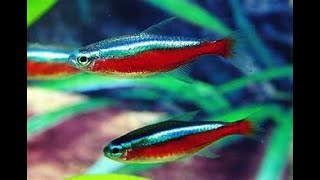
Fish disease case nr. 363 Wild Cardinal tetra, Paracheirodon, with worm and bacterial infection
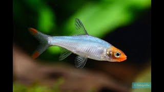
Fish disease case nr. 362 Sawbwa with gill flukes, Nematodes Capillaria and bacterial infection
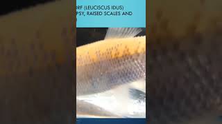
Sick Golden Orf with Dropsy. See link complete video #fishdisease #GeraldBassleer
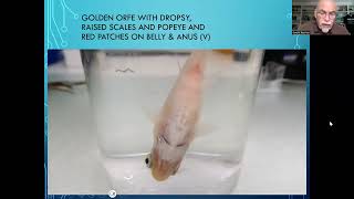
Fish disease case nr. 361 Golden Orfe with dropsy or ascites due to bacterial infection
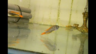
Fish disease case nr. 360 Apistogramma with 3 different parasites and tubercles in the organs