This Is What Filler Looks Like On Ultrasound

21 Oct 2026 · 1 min
1:04

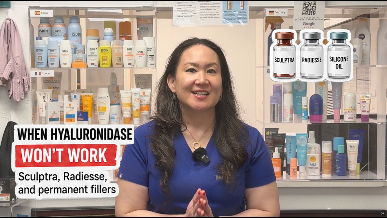
In this brief educational video, board‑certified dermatologist Dr. Jane Yoo demonstrates how different dermal fillers appear on ultrasound and explains why identifying the filler type, depth, and exact location is essential before using hyaluronidase. She describes the typical ultrasound patterns: hyaluronic acid appears dark (anechoic to hypoechoic), calcium hydroxyapatite (Radiesse) appears bright with acoustic shadowing, and silicone shows a snowstorm pattern. Dr. Yoo emphasizes that these imaging cues help clinicians confirm the presence of filler, distinguish the product, and avoid ineffective hyaluronidase treatments, since the enzyme only dissolves hyaluronic acid.

Chapters

More from Jane

7:51
youtube.com

When Filler CAN'T Be Dissolved: Sculptra, Radiesse & Permanent Fillers

1d ago
7:13
youtube.com

What Botox Actually Does To Your Face | Dermatologist Explains

1w ago
7:40
youtube.com

Is Dissolving Filler Safe? Side Effects & Risks, Explained

1w ago
7:03
youtube.com

Does Dissolving Filler Cause Sagging? A Dermatologist Explains

2w ago
1:10
youtube.com

Does Hyaluronidase Dissolve Your Own Tissue?

2w ago
7:15
youtube.com

"Hyaluronidase Ruined My Face" | A Dermatologist Responds

2w ago
1:02
youtube.com

Chin Filler Migrates Down, Not Out

2w ago
1:03
youtube.com

Why Your Under Eyes Puff Up After Cheek Filler

3w ago
0:55
youtube.com

Hyaluronidase Doesn't Work On Every Filler

3w ago
0:55
youtube.com

The Tongue Test for Lip Filler Migration

3w ago
6:51
youtube.com

Filler Migration by Area: Chin, Cheek, Nose, Jaw & Temple

3w ago
See all content