Clinical Series: How to See a Pinched Nerve (Foraminal Stenosis) on MRI
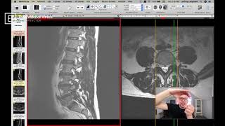
This is a narrated YouTube clinical tutorial by Dr. Jeff Langmaid explaining how a pinched nerve (foraminal stenosis) appears on lumbar MRI. The speaker demonstrates sagittal and axial T2-weighted images, identifies disc bulges/herniations intruding into the foraminal canals, and connects specific levels (e.g., L4–L5, L5–S1) to typical patterns of leg pain (dermatomes). The video aims to help patients recognize imaging signs of nerve compression and to guide clinical decision-making.
Chapters
Hi, my name is Dr. Jeff Langmaid and today we are going to review exactly what a pinched nerve looks like on an mri. If you have suffered with a pinched nerve or what is commonly called foraminal stenosis, you know the challenges that can create in your life. Many people that have a pinched or a compressed nerve experience not only pain right where it's occurring, but pain that also travels down their arm if it's pinched in the neck, or down your leg if you have a pinch or compression in your low back. So today we're going to take a look at what that actually looks like on an mri. We're going to evaluate it, take a peek and give you the best information so you can know what you're looking at, what challenges you are up against, and also hopefully how to make a great decision for your health to find relief. So without further ado, here is what an MRI looks like when there is a pinched nerve present.
Here we are within Ocirx, so I use a Mac and I prefer the image viewer of Ocirics to do so. Ocirx has a free book and I believe Radiant, if you are a PC person is the equivalent on a PC. But this is a lumbar spine, a lumbar MRI and a couple things to note right off the bat. So on the left hand side of the screen, that is a side view, a lateral view or a sagittal view, they all mean the same thing, as if the person is facing sideways and the picture was taken from the side. So up in here is to tummy or stomach, we have the buttocks going down here and the belt or the waistline would be right around here somewhere. And on the right hand side of the screen is the axial view. Now keep in mind with an axial view on the right hand side of the screen, it is looking up, not down. So left and right side are reversed. Always important to keep that in mind when looking and reviewing MRIs. I have T2 weighted images up on my screen and and the big gray squares, as you well know, are the vertebra or the bones and the dark spaces in between are the discs. That bright white stripe down the backside is ultimately the spinal cord or the thecal sac. So that is the highway where all the nerves are traversing up and down. You can see it right there, that bright white stripe. So in an ideal world, as you know, what we like to see are the discs directly hang out directly underneath the vertebra or the bones. They should be nice and flat on one side and on the other side. And as we look here, we can notice this segment for instance, is very flat on the backside. That's great. This is quite flat as well. But when we get down here, you can see dark material has extended posteriorly or back towards where the nerves reside. And that is very valuable real estate. The amount of space where the spinal cord or the cauda equina go through is only at maximum about 10 millimeters in width or dimension. So even just 2 millimeters of a disc poking out, whether it's a bulge or herniation or an extrusion, takes up 20% of that space. And the foraminal canals, or where the nerves travel out the side where we'll see the pinched nerve, those holes, those canals are even smaller. So here we have the disc poking out the backside here. As we go down towards the next level, we can see a little hump out the backside there, a disc bulge there. And what do you know, we have one here as well. So there are a couple different levels at first look that indicate we might have a problem where the nerve is traversing out. So on the low back in the lumbar spine, we're going to. One of the great things about the lumbar spine is you can stay on your side view or your sagittal view and just go out towards the side and you will notice as it go out towards the side. Let me get the right slice here for us to look at. As you go out towards the side, you can actually visualize those huge holes and canals where the nerves come out, the foraminal canals. I'm circling one on the screen right now. Look at that big white space. Look how tall it is and look how wide it is. That is awesome. If we go up to the next level, look how tall that white space is and look how wide it is. That is a fantastic looking foraminal canal. When we go down here, we see again, look how tall that space is and look how wide it is. And the next one. Alright, that starts to look a little bit hourglass or a little bit like a keyhole. And why is that? Well, we see a little bit of that dark disc post poking out into the bottom. It's not really compromised, but it is less than perfect. Right. And if we go down one more level. Oh gosh, that white space is starting to look a lot thinner or there is not nearly as much white space here as we had way up at the top here. And now we go down to the our trouble level. Look at that. There is just Absolutely no space for that nerve to go out. That's actually the nerve right there coming out at us. So there is a very limited amount of room where the nerve wants to travel out. And why is that? Well, half of the hole where the nerve travels out is the top bone, half the hole is the bottom bone. And if the disc is poking back into the hole, it can take up a lot of that real estate and push the nerve towards the top. And if the nerve gets jammed and pushed up in the top aspect of the bone and has the disc below it, it ends up sandwiched between the two, causing, drum roll, a pinched nerve, or what we would typically call foraminal stenosis. Now, when we see foraminal stenosis, specifically in the lumbar spine, our expectations of what a patient is going through is pain traversing down into the thighs, down into the calf, down into the foot. It is ultimately the textbook definition of a pinched nerve. And if the nerve is compressed at the site where it comes out, that tells you where the pain is going to go to. So I always explain it like power lines. Every different level that you see here, every different pathway where a nerve travels out is like a different power line going to your neighborhood. They all go to different houses. So you could identify based upon where the power line is going to go to what house it may affect. And we know, we call that dermatome. So we know if, let's say L4, L5 is compromised, that level, many common level, for many people, that tells us that it's very likely to be pain down into the calf. We know if L5S1 is compromised, there could be pain all the way to the big toe. If it's L3, L4, it might be more on the front of the thighs. Of course, these overlap a little bit, but there are some clinical hallmarks of the pain that you may be experiencing and what level it may be correlative to in your spine. So you could hopefully clearly see this is a severely pinched canal compared to what you have as far as that big, bright white space up above. Now, if we go to the other side of the screen and we examine it from the axial view, I click over there. You can see our axial view is the cut you in half view. It's as if you sliced somebody and then looked up. So this is the well where we're at now. This green line tells us where we're at. If I move it a little bit up, we are in the bone. So that is the vertebra or the bone. This What I'm circling right here on the right hand side of the screen. This is muscle on the backside. Muscle on the backside. This is the spinous process. And this big white space is the central canal or spinal cord. This is actually the thecal sac. All of those little black ants as I call them, those are the individual nerve roots that are traveling down. So that is the lay of the land. And, and if we go down into where that pinched nerve is on the foraminal canal, in the foraminal canal here is what we will be looking at. That is a disc right there. This is the central canal. It is not perfect, but that's not our focus today. It's a little small. This is a facet joint or a little knuckle on the backside of the spine. You may have heard about the facet joints. This is the other one. And this is the backside of the bone going up. Now what I want you to appreciate is this little gray or white area here. It doesn't look great, but if you appreciate the fact that there actually is white space where my mouse is here and the fact that there is no white space, it is completely dark on that side. This area that I'm pointing to right here is our problem area on this side of the screen right here. So ultimately what we're visualing on this side is just a different view of the same thing. The disc has bulged or herniated and it has pushed in to that very valuable real estate where the nerve wants to travel through. And when that happens, we call that foraminal stenosis. You may call it a pinched nerve, but quite often that can result in pain that travels down an arm if it's a pinched nerve in the neck or, or the legs and feet, if it's a pinched nerve in your low back. And that is how you visualize it on an mri. So I hope this was very valuable. If you have any follow up questions, please feel free to leave them down below. I'd be happy to answer any questions or if there are any other items that you would like me to show and take a look at on mri, I'd be happy to do so. Leave that down in the comments as well. I hope you have a fantastic day and I look forward to talking with you soon. Bye bye.