Live MRI Review: Complex Lumbar Spine Fusion and Radicular Pain
Dr. Jeff Langmaid reviews MRI scans of an elderly patient with a three-level lumbar fusion and new radicular pain. The images show a postsurgical seroma, disc extrusions above the fusion, and severe central canal stenosis at L2-L3 causing neurogenic claudication. He discusses adjacent-segment disease and conservative treatment options like anti-inflammatories and flexion-distraction.
Chapters
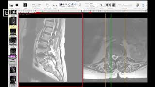
Hey everybody, and welcome to the Big Picture. This is a weekly radiology review. I'm Dr. Jeff Langmaid with the Evidence Based Chiropractor. And today I'm going to break down Share My Screen and we're going to take a look together at a complex multilevel lumbar spine fusion. This patient came in to see me quite a few years ago. He was in his late 70s, had reported on his history previous back surgery, and he had all of the same challenges as he reported. They were even worse now than they were back then. We're going to take a look through that MRI together. Comment below with your findings as we start to dive in. The goal of the Big Picture is to get you more and more comfortable seeing complex MRIs and maybe some not so complex MRIs as well. Little bit here, a little bit there. That builds your confidence and will get you much, much, much more more comfortable reading MRIs. As always, I give the caveat. Please continue to rely on your neuroradiologist for all of those organic complaints. But I want you to be able to read comfortably and cleanly as you go through. So let me share my screen. It looks like I might have got stuck there. And here we go. So what we have here is a lumbar style. So on the left hand side of the screen you can see we have our lateral cuts. On the left are the laterals. On the right hand side of the screen we have our axial cuts. So right hand side of the screen we have the axial cuts. So T2 weighted images. I always like to bring my laterals or sagittals on the left and then I like to bring my axials on the right hand side of the screen. So let's start over on the left. What we can notice right here is we are in a center cut, right? So my scout line tells me I am right in the center. And I can see the vertebral bodies. You can see already down here, looks like 5, 1, we have a fused disc space there. At 4, 5 we have a potentially fused disc space and a slight offset, at least a grade one spondylolisthesis there. And then up at 3, 4 we can see we have end plate changes. So that disc where the disc meets the vertebra, the end plate, those are modic changes. That looks pretty nasty because it just looks blurry and just unclean. When we get up to two, three, we see a lot of the same end plate change. We see disc hanging out the backside. And as we get up towards 1, 2, we see more of the same. The disc. Excuse me. The end plates do look a little cleaner, but we can see a disc hanging out the backside all the way up there. And finally, at the top of that lumbar spine we, we see a relatively normal disk and disk space. So we have L5, last fully formed disk space at L5, S1. So we have S1 small in S2 down here. L4, L5, L4 and 5. We have a spondylolisthesis previous. It looks like that disk space has been fused. At 3, 4, we see challenge end plate change and disk hanging out the backside. Two, three, a lot of the same. One, two, some disc coming out posteriorly. And one, two. Excuse me. And 12, one looks pretty good. Down the backside we see cauda equina. So right where that thoracic spine typically meets the lumbar spine is where the spinal cord breaks up to become the cauda equina. And we should see a three lane highway for where our CSF is white, the spinal cord or cauda equina, dark and then white behind it. The white enables space, right? Cerebrospinal fluid gives space. And we can see at 5, 4, 3, 2. At L2. At L2, L3, we have a potential major pinch point going on here. We have a three lane highway becoming a sidewalk. And that is because we see facet potentially poking out the backside posterior to anterior. And we see disc poking out the front side anterior to posterior. That's making a very, very tight space. Additionally, when we get down towards L5 posterior to L5, we see a nice big space for the cauda equina to go through. A lot of big space for the csf. But we also see an additional white spot, so to speak, a pool of white. Behind that posterior to where we'd anticipate our spinal canal, we see an additional piece of white. What could that be? Well, very often, especially in a previous surgery case, that is a fluid collection. Fluid collections are unbelievably common post surgery, but typically they disperse over the first few weeks and or months. However, some become encapsulated. The fluid collection becomes encapsulated, that means it's not going anywhere. And that is typically called a seroma. So let's look side to side a little bit on our sagittals here. We'll go out towards the side and lo and behold, what do we see? We see the hardware come into play, right? So we can see at 5:1, we see some screws at 4, 5, we see some screws at 3, 4. So we see a 3 IC, a 3 level fusion going on. We see a 3 level fusion going on posteriorly instrumented lumbar spinal fusion. Now, if we go towards the other side, what do we see? Lo and behold, we see the same. So we see there is bilateral instrumentation. So there are screws coming in the left and the right side all throughout that lower lumbar spine area. And that makes sense with what we see with the fluid collection. And it makes sense whenever you see a big pinch point, a large compression spinal stenosis, foraminal stenosis above an area that typically is indicative of adjacent segment disease. So that's typically indicative of an area that has undergone additional stress and strain as a result of effusion happening below. And that is exactly what we see up there. Now let's head over to our axials and to get a better interpretation of what's going on with the spinal canal, we go over towards our axials. We are going to bring it down all the way, all the way down towards our L5S1 junction point. And we can see on our axials we have plenty of room. I like the amount of space that we're seeing right here and rightfully so. They've been decompressed. Right? This person's had a surgery to give them space. It looks pretty good. As we continue on up, what do we see? Ha. We start to see our fluid collection. Not only do we see our screws come in side to side, but we see that large scale fluid collection towards the backside. The fluid collection that we see right here, a big white space behind your spinal canal on the axials or below. I guess you should always rectify against your sagittals. And it's very clear that we are looking at the same fluid collection. Left hand side of the screen and right hand side of the screen there. We see the screws coming in. This person does look fully decompressed. We can see, as I call the little ants, are the nerves traveling down, the individual nerve roots traveling down. They look pretty darn good. Now let's continue on up. We continue towards the next disc space. Okay, we see it got a little blurry, but bottom line is this still has good A to P diameter and we're still seeing our fluid collection on the backside. So we still have fluid collection. It is not near, as you can see here, it is not near to the skin. So this is not necessarily at risk. Sometimes I've seen fluid collections directly under the skin and that's a, that's a high risk of potential infection. It's a high risk of broaching it and causing big time problems in an outpatient setting, for sure. This, this is a deep fluid collection directly posterior to where surgical intervention and instrumentation occurred. And as we continue to go up to the next level now, we start to see some challenges. Right. What happened to our spinal canal? We have our disc up top. I'm circling the disc. That should be clear. And we have our facets on the backside, our left facet here and our right facet here. But we can see we have supreme, supreme triangulation of the canal. Watch as I bring our green line down just a touch. Look at how that pops open to bright white. Plenty of CSF here. And watch this bright white area become significantly darker as we get up towards that disc space. This person still has some breathing room there, but there is triangulation of the canal. There's a little bit of facet hypertrophy going on. There's some fluid. I see a little bit of fluid in the facet right here. But watch when we go up a little bit. Very commonly, this is what occurs. We see that pop open when you get by the disc space. So we are now above that disc space. We can clearly see that the spinal canal has popped open. So there is room again. Now, we talked earlier. We are headed into a potentially treacherous area right here. So watch what happens to our spinal canal as we continue to go cephalad. We are looking good. We already start to see a disc, right? Bright white, love it. Nice and big. We start to get up here. I see a disk start to come in. So we have an extruded disc going on at this level. So let's say it's five. Let's call this five, four, three, two. At two, three. We definitely have an extruded disc. That is disc material. And we have not got to the inferior portion of the end plate. So that is sagging down the backside. And as we continue to go up. Yeah, now we see some significant pressure, some significant compression. We see facet fluid side to side. The white stripes in the facets tell us there's fluid and inflammatory response there. We see ligamentum flavum. This is exceptionally wide, so to speak. Ligamentum flavum is very hypertrophic and it has caused a supreme compression on the spinal cord and spinal canal at this level as we continue to ascend up. Look at that. One sec, one slice. That's only a couple millimeters. And look how much that opened up. Watch the difference between that. That is tight, tight, tight as the Dickens and one up and look how that pops open. So if we calling this L5, L4, L3, L2, we have a significant spinal cord compression going on at L2, L3. That would be a very good reason for the neurogenic claudication that this patient was experiencing. He was experiencing weakness in the legs, pain in the legs, challenges when walking, and L2, L3 seems to be a significant contributing factor to those challenges. Now, as far as axial pain is concerned, well, number one, he's had a very large multi level fusion, so that can always cause downstream axial pain. Additionally, we have a fluid collection. That fluid is not supposed to be there. Now, depending upon how post surgical he is, if he is one year post surgical, I would not expect that fluid collection to be there. If he's two months post surgical, this still might have an opportunity to dissipate. That's where it comes down to be very key when you're asking your patients that have had previous surgery, how long has it been since that surgery? Again, fluid collections are very normal. They form in many, many surgeries of the spine. I've seen tons in the lumbar spine. The good news is, is that most of the time those fluid collections dissipate over time. It's only if they become encapsulated or become a seroma that they could stick around ad infinum for a very, very long time. So this was a quick insight into a complex multi level fusion which we had everything in the kitchen sink going on. We have degenerative change, we have modic end plate changes, we have extruded discs, we have previous posterior instrumented hardware. We have a fluid collection. We had it just about all going on in this image. Bottom line is this patient foraminal canals look pretty good side to side. He's a little bit snug in some of the foraminal canals above the previous fusion, there's extruded disc above the previous fusion at multi levels and but specifically with our naming conventions, we used at 5, 4, 3, 2. At L2, L3, a predominant factor and predominant finding in this individual's case is significant spinal cord compression. At L2, L3. There is no reason based upon these images that you would be unable to treat that area conservatively and see if you can get gains. Quite often. For people with significant spinal cord compression, you're going to want to, you know, make sure that they're in tune and in communication with their other healthcare providers, including their surgeon or neurologist. However, there's no reason why you shouldn't be able to get in there and see if gaining an additional millimeter or two by decreasing inflammation, by increasing motion. Potentially this is a great case for flexion distraction. Getting any opening on that spinal cord and canal that you can quite often can provide significant relief for that individual, even if only temporary. That is still a huge step in the right direction, especially for somebody who's a little bit aged at this point in time and has undergone multi level fusion in the past. Staying as conservative as possible, barring any red flags, is a great way to go. So hopefully that gave a little bit more additional insight into a complex lumbar multilevel fusion. This has been the big picture. My name is Dr. Jeff Langmaid. Have a great day in practice.