LIVE MRI Review of a Complex Cervical Fusion: Key Evaluation Points
Dr. Jeff Langmaid reviews an MRI of an elderly patient with multilevel cervical fusion, highlighting how to spot adjacent-segment disease above the fusion site where severe spinal cord compression often develops. He walks through the imaging workflow: sagittal views first, then axial slices to assess CSF space and canal stenosis, emphasizing that direct image review catches details radiology reports miss.
Chapters
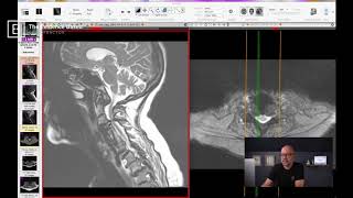
Hi, My name is Dr. Jeff Langmaid with the Evidence Based Chiropractor and today I want to break down a complex multi level cervical spine fusion on mri. We probably have all seen patients who have had a large scale fusion in the past and it can be scary, it can be intimidating, and a lot of times there can just be a lot of fear for what a patient could or should do next. If you are their doc at that moment in time and they want you to take a look at those images, I want to go through some of the key findings that I have found in the past. This is a past patient of mine and also just identify some landmarks so you can feel comfortable and confident having these conversations with your patients and helping guide them to the next step, wherever that may be. So let me click over and share my screen and then we will get started. Started. Let's see here. Cool. So here we go. Up on the screen is a patient, this patient is or was in their early 80s. So we can see we are looking at the cervical spine. I'll kind of bring my cursor over here. On the left hand side of the screen we have the lateral view. So the lateral or the sagittal view is what we refer to on the left side of the screen. On the right side of the screen over here, where you see my cursor go, is where we have our axial views. So again, we have our lateral or our sagittal on the left and we have our axial, which are the slices. As if you took a slice like this and looked up those cross sections. Those are on the right. So on the left, just identify some landmarks real quick. We have the brain up top. You can clearly see that. You follow my cursor here we have the chin, the lips and the chin. So this is a side picture, right? A lateral. If I move over a little bit, we want to get in the center, we can see that skunk stripe is the spinal cord going down the backside. So we can see brain up top, spinal cord coming down the backside. Directly in front of that spinal cord are our vertebral bodies. So those big gray squares are the bones, as you can see here where my cursor goes. And in between those vertebral bodies, in between the bones are the discs. That's the dark spacing across the middle here. Now one thing to note is right in this area, you might be saying, gosh, those do not look like normal discs. And you would be right. This is exactly that area where this patient has had a large multilevel cervical spine fusion. My, my opinion, it actually looks like they had a corpectomy or potentially this fusion was done many, many years ago. I'm not exactly sure. But you can clearly see there is a lot of changes. There's a lot of bone that is all fused together right here. There isn't too much disc material. And if we slide over, we see these dark lines going up and down. They almost look like a strut. So struts are used in large multiscale or multi level procedures and those tend to be used in corpectomies. This person looks like they still have some bone left. So a little bit confusing. I'd love to look at an X ray of this person to get an absolute idea of the construct itself. But you can see above this. Here's where we start to get into some challenges. Well, let's identify our spinal cord. Right behind that fusion is the spinal cord area. We can see white, dark and white. The white on the front or the anterior side and the white on the posterior side. That is the cerebrospinal fluid. That's the cushion around the dark stripe in the middle, which is the spinal cord itself. Now this has plenty of room. You can see my cursor going anterior to posterior, back and forth. Right here things look pretty good. But when we get up above, wow, it looks like that three lane highway right here of white, dark and white becomes a sidewalk. There is absolutely no white on the front. There is no white on the back, which means there's no breathing room. There's a significant amount of compression going on above that previous fusion site. We kind of cruise over here. This would be where C1 is. We see C2 and C3. So this person has a C3, C4. Significant, significant spinal cord compression. Now here's one of the key findings and takeaways. Anytime that you see a single level or a multi level fusion, one thing you always want to take a look at is the level above. Adjacent segment disease can take place in a majority of cases where the spinal fusion takes place. And that is because all of these individual segments, the disc and the bones, are designed to work independently and to share the load. But when a segment or multiple segments get fused together, obviously they don't share the load at that point in time. So the level above and below, but specifically above tends to take a mass. It works overtime. It takes a massive amount of extra stress, a massive amount of extra load. And that can lead to advanced degenerative changes. It can lead to breakdown of that segment. It can Ultimately lead to, as we see here in this person's case, some advanced spinal stenosis at the 1, 2, 3, at the C3, C4 level, where this individuals has a C4, 5, 5, 6, and maybe even 6, 7. I'll take a closer look. Multi level fusion below that. So that's kind of taking a very quick look at our lateral or our sagittal view. That's probably what most docs feel comfortable doing. But really what we want to get a great idea of is the other side of the coin, which is the right side of the screen. Over here, Our right side of the screen is our axial cuts. So we can see right here we are in our fusion. Right. Our cut on the right side of the screen is just the slice of where our green is on the left. Right. It tells us our positioning. So we can see the disc is up here, disc or bone, depending upon where we're at in the slice. And the big white circle on the backside is our spinal cord. So we see our spinal cord here. We see the white is the cerebral spinal fluid, just as we saw on the other side of the screen. And the dark is the bundle of nerves, the cord itself. So as we kind of move around the fusion, I'm feeling pretty good. This person, you know, there's a lot of white. There's some nice dark in the middle. That tells me that there's some space. I could nitpick apart a little bit of stenosis within the fusion. But watch what happens to this huge white circle as I bring our green line up above. Watch what happens to all this space. We're looking good. We're looking good. We're looking good. Oh, my goodness. We saw all of this disappear. Where did our white breathing room, where did that CSF go? It is being compressed and pushed out of there because we have disc, bone and ligament. Disc kind of going from anterior to posterior. And then we have ligament and bone pushing posterior to anterior and the spinal cord, the spinal canal is being crunched in the middle of that. Watch when I continue to cruise. We're right in there right now. There is hardly any white. And I go up above. Oh, my goodness. Breathing room. There we go. So just like that, watch right here where my cursor is on the right side of the screen. We're right above where the area of challenge is. We are in it right there. It gets dark, we stay in it and we pop back open, lo and behold, right here. And things look open and clear. My point of this is when somebody has a large scale Fusion. There can be challenges inside of a previous fusion site. I have seen it time and time again. But they're also keep a keen eye on the areas up above. Now, in this individual's case, being 80 years old, is there going to be another surgery in their future? That depends a lot on the symptomatology and what's going on. Clearly this individual cervical spine has a lot of issues. When we go down below, if I'm back on the left hand side of the screen, we go down below the fusion site and we can see degenerative changes in the discs at near every level. We can see disc bulges, those little dark humps pushing out the backside. We can see bulges or herniations at multiple levels. We see modic changes on the end plates. There are a lot of findings. I mean, you could probably just name about anything that you've ever seen on an MRI with this individual. There's foraminal stenosis, there's lateral recess stenosis, their central stenosis and more. There's a lot going on on this individual's mri. But I just want to show an example of a large scale fusion, what it looks like on an MRI. This is the T2 weighted images of that MRI, in case you were curious. I'm looking at the T2 weighted images and also just show you what happens above because above a multi level fusion, for every level that you add on to a fusion, it dramatically increases and there's like a multiplying effect, the amount of stress and load that it places on the first free level above that fusion. So keeping a very keen eye and just looking. A lot of docs feel comfortable and or patient, patients even feel comfortable if they pop in their MRI disc to look at the side view because you can kind of see where somebody's nose and lips and chin are and you can see where the brain is. It's a pretty easy view to look at and see. But keep a keen eye on the other side on your axials, because your axials are going to tell you the amount of compression. You can see the fact on the left hand side, on your laterals or sagittals that there is compression, but your axials on the right show you the amount of the compression. In this individual's case, he has severe spinal cord stenosis above the previous fusion site. These are these large. On the right hand side of the screen we see these large dark areas. This is massive facet hypertrophy and facet disease. Probably because those facets have also undergone a lot of Extra stress and strain due to the fusion below. So we see facet disease, we see foraminal encroachment or foraminal stenosis where the nerve is trying to go out the left and the right side and service the, you know, service the shoulders, arms and hands when we go lower. And we also clearly see there is a significant amount of central cord stenosis or compression on an individual like this. Some of my questions immediately would be regarding balance. I want to make sure, or at least question whether or not there's any damage going on on the spinal cord itself. Now they might have had that previously and that's where they had a multi level fusion. But I'm going to be curious to know about potential balance issues because that could lead me towards a myelomalacia type thinking at which point I would want a neuro consult immediately. I'm going to be asking about quality of life, I'm going to be asking about headaches, I'm going to be asking about, you know, basically stenosis type issues, upper motor neuron issues, are they able to ambulate, are they having difficulty ambulating? What's going on with the strength in their arms, hands and shoulders. I'm going to want to get a eye on all of that from a subjective asking the patient point of view, but also from some objective tests in my office because clearly this individual has a lot going on. So anytime that you have somebody comes into your practice with a large multi level surgical fusion, I like to set my images up. I look at the T2s on the left hand side of the screen. I always drag my lateral or my sagittal on the right hand side of the screen. I always drag over my axials or my cross sections and from there going to take a look within that fusion site, get a good idea of what's going on with the spinal cord. Again, there's some less than perfect things going on foraminally with this individual in their previous fusion site. But the spinal cord by and large looks pretty good, especially when compared to that level above. So I hope this gives a good insight into a pretty complex case, a pretty complex surgical case. If you have any questions, feel free to leave them down below. I'd be happy to answer. And also just let me know and comment if you'd like to see more of these complex cases. Cervical, thoracic and or lumbar post cases, let me know. I'm happy to review them. And anything that you'd specifically like me to go over, just let me know. I have a large quantity of a large library of MRIs. They've all been sanitized, but they are all fantastic to look at and get a better idea of what's going on with patients in my queue. So when the patient walks in your front door, you can have a better opportunity to explain what's going on and guide them without only having to rely on just the reports. So that's a practical assessment of a complex cervical spine fusion. I hope you have a great day and I will talk to you soon. Bye bye.