Lumbar Fusion Surgery Gone Wrong: A Live MRI Review
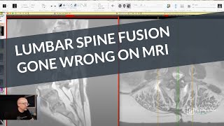
Dr. Jeff Langmaid reviews an MRI scan of a patient after multilevel lumbar spine fusion, examining what went wrong with the procedure and the complications visible in the imaging.
Chapters
Hello and welcome. My name is Dr. Jeff Langmaid with the Evidence Based Chiropractor and you are tuning into a live MRI review. I'm going to share my screen in just a minute and we're going to go through an MRI of a patient who had a multilevel lumbar spine fusion. Yes, it was a big procedure. Yes, there were some residual challenges and that is why I saw the patient in preparation practice. But I want to show you exactly what that looks like on the MRI and also break down some of the challenges that this patient continued to have. Now, you probably know if you have long standing low back pain and a significant pinch or stenosis on the spinal cord and or your spine is unstable, those are all indications for potentially having a fusion surgery. The big challenge with the fusion surgery is it is risky, it takes a long time and it ultimately compromises your spine forever. We are not made to have rods and screws in our spine. Hey, there are those rare occasions when it is a godsend for individuals to have it done. But what we've seen time and time again is that over these last 10 years, the incidence rate of spinal fusions has dramatically went up and it has not really impacted the results and outcomes that people are receiving. What I always hear from surgeons, sort of when we're having lunch, having dinner, is about a third of their patients do a little bit worse, about a third do better, and about a third remain the same. Those odds are not exactly great. Now again, there are red flag situations where a fusion is an emergency surgery and unbelievably necessary. But. But elective surgery is different matter altogether. We're going to take a look together through exactly what this looks like on an MRI and some things to look out for just clinically. So let's share screen and I'm going to head on over and we'll take a look together. So here we go with this patient. So this is a lumbar MRI image. On the left hand side of the screen are the side view. That's what we call the sagittal view or lateral view. It's like you took a picture of me just as the camera is looking at me this way. That's your left hand side of the screen. On the right hand side of the screen is what we call the axial view. It's as if you took a slice like this and then looked up the only difference. So it is a cut and look left and right are reversed because you're looking up instead of down. But that's what we have on the screen here. I'm going to start on the left hand side of the screen. The yellow is showing us. Those yellow lines on your screen are showing you exactly how far up and how far down the scan goes. And the green line is what we refer to as the scout line. It showcases to you where you are in space. So the big gray squares here are the bones. Your big gray squares are the bones. Those are the vertebral bodies. This is L5 all the way down here. This is L5, that's S1. Above L5 is L4, I'm circling that right now. L3, L2 and L1. So the big gray squares are the bones and the discs go in between the dark material. In between those great big gray squares or the vertebra are the disks which they are supposed to be there. That white area going all the way down. If you see me tracing it all the way down the posterior or backside of the bones and discs, that is the spinal cord or the spinal canal. And it becomes the cauda equina as we get down towards that lower lumbar. We're looking at a lower low back right here. So in an ideal world, I'm going to go up to L5, 4, 3, 2, 1, L1. You can see this, what I'm outlining right here. This is a nice square, right? This bone is square. The vertebra is square. It's nice and gray. It's clear around all of the edges. That is normal. That is a good looking vertebra. Now one thing that you will notice as we go down is these things start to look sloppy and different, right? That disc, instead of residing nice and underneath the bone, we see multiple areas where the disc is poking out the back side. Look at those, I call them like a beak, right? Poking right out the backside and or out the front. So we have degenerative changes at L5, S1, L4, L5, L3, L2, L1, L2 and even T12, L1. So we have a lot of degenerative changes throughout the spine. The other thing that you might be taking a look at and saying, Gosh, at L4, L5, we're on the left hand side of the screen. That disc looks a lot different than the others. And that is because it's not. It's no longer a disc. That is where a previous fusion. Watch. When I scroll to the side a little bit, bam. You can see all of those rods and screws, these big black areas going in. Those are rods and screws. That is A multi level spinal fusion, I believe it's L4, L5, S1, multi level spinal fusion, L4, 5 and 1 have all been fused. Look at that hardware going in. So that is why between L4, L5, that disc space right there, between L5, S1, that disc space right there, that's why it looks so different, is because the hardware is around. And when they do a fusion, ultimately you are kind of put out. Obviously you're under sd, you're cut open. And they remove as much of the disc as possible and put a spacer in there, an implant or a spacer in where that disc used to be. Some of the disc remains, of course, but they take out as much of this as they can put in a spacer and that sort of stabilizes everything. And then they put the rods and screws in to help support that. So that's what we are seeing right there. As we go towards the sides are a big mess of hard Hardware at L4, L5 and S1. Now, one other thing you might note and see very quickly on this image on the left hand side is there is this big weird pocket of white stuff hanging out behind where the spinal canal is. That is a large, large fluid collection. So that is a fluid collection that has happened as a result of the surgery. Now, in almost every single lumbar, or cervical for that matter, almost in every single spine fusion, there is fluid that gets collected in the area. However, that fluid is expected to dissipate throughout time. So nearly every single surgery has a fluid collection right afterward. But that should dissipate throughout time unless it becomes encapsulated, at which point we call it a seroma. And that means it is not going anywhere. So fluid collections by and large don't cause a problem at every time, but they can cause a problem on occasion depending upon exactly where they're located. Are they near the nerve, are they in and around the hardware? All of those factors matter on whether or not that fluid collection is ultimately going to cause a problem or a challenge. So important to keep that in mind. Now let's go over to the right hand side of the screen where those axial views you can see on the left hand side. It's going down, right? So it tells us where we are. Right now I'm right at that L5, S1. You can see right there between L5 and S1 kind of in that this disk space. And what do we have? Well, this is the spinal canal. So what I'm circling right here is nice and big open and wide. That what we're looking at is the space right here that I'm outlining on now, the left hand side. So that is where we are at. Now let's go up a little bit. What you'll notice is look at all the two white spaces appear right when we go down on the right hand side of the screen. One white circle, spinal canal up a little bit and we see that second white area. That second white area is the fluid collection. So again we go back over to the left hand side of the screen. We can see right now. Oh boy. We're right there. We're right where that fluid collection is in back of the spinal canal. We see. See that right here. Now the spinal canal looks wide open and that's fantastic. You can see how tall it is here and how wide it is across. It looks fantastic. To Continue up to L4, L5. You can see that again, fluid collection sitting right behind, right next to that spinal canal. But it looks great overall. As we continue to go up. Oh, now we start to see some challenges. That big open circle, that was the spinal canal. Now it's become a very small triangle. Look at this white triangle on the right hand side of the screen. That is stenotic. It's compressed. If we go down, watch it open up to be that huge big circle down at the next level. As I'm circling right here. Or if we go up, you can see it crunches right down into a triangle. So there is definitely some stenosis or more compression at L3, L4 right above the fusion in this individual. Let's keep on going up. As we go up towards L2, L3. Oh boy. We see severely stenotic. Look at that little tiny area that I'm highlighting that is left of the spinal canal or spinal cord. Even compared to the level below. Level below. That looks a lot bigger and healthier to me. We could measure, but that looks like I can just see it visually. That is much more healthy. It's probably moderately stenotic at L3, L4, but there is severe spinal cord stenosis at L2. L3. Additionally, we see fluid in the facets. Those little white streaks that's fluid in the facet joints. And we see that big black V right here is a ligamentum flavum. It's a ligament that sits inside of the spinal canal that's very thick and hypertrophic. Is causing a lot of, a lot of challenges for this individual. So really, really stenotic as we get up towards L1, L2. Look at that. Looking great. We see a little bit of a disc bulge right there but look how wide open that spinal canal is compared to what we had down below and how compressed that was. So this individual, they have probably had previous back pain for a long period of time because we can clearly see that this individual had a multi level fusion. Now with that fusion unfortunately it's L4 L5S1. With that fusion it resulted in a pretty large fluid collection which might not be the source of the issue but we can see up above now there's that cascade effect start to stabilize and secure infused one level it puts additional stress and strain on the levels above. That's exactly what's happening right here. We see an L3, L4, 4 and more importantly L2 L3 a huge pinch or compression on the spinal cord. So I hope this was helpful. If you have any questions whatsoever, feel free to leave them down in the comments. I'll keep an eye down there, make it a great day and I'll see you soon.