Hydroxy Radical Protein Footprinting (HRPF) and its relevance in Drug Development
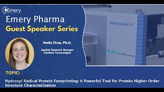
Recorded talk by Dr. Emily Shea (Applied Research Manager, GenX Technologies) introducing hydroxy radical protein footprinting (HRPF/FPOP) as a mass-spectrometry based structural biology method. The presentation compares low-resolution (biophysical) and high-resolution (X-ray, cryo-EM, NMR) approaches, positions mass-spec footprinting (HDX, chemical crosslinking, HRPF) as a mid-to-high information, solution-state technique, and details GenX’s laser-free FOX (flash oxidation) system that photolyzes hydrogen peroxide and uses an adenine dosimeter. Applications shown: epitope/paratope mapping for TNFα–adalimumab, detecting aggregation interfaces and formulation effects, and mapping conformational changes in ABL kinase with allosteric inhibitor GNF5. The talk ends with audience Q&A on quenching, aggregation mitigation, and comparability/stability uses.
Chapters
Good afternoon everyone. Thanks so much for joining us today. My name is Neeku Madhavian. I am the Director of Corporate and Business Development at Emory Pharma. We're very excited to announce that we have established a co marketing agreement with genex Technologies and we've recently press releases as well. I'm pleased to introduce Dr. Emily Shea. She is the Applied Research Manager at GenX Technologies. She earned her Ph.D. under Professor Lisa Jones at the University of Maryland in Baltimore. Over the past three years at GenX, she has made strides in development of HRPFs. You're probably wondering what that stands for. I'm not going to get into the details because Emily will be sharing more about that. Today's talk will also cover how to do high order structure analysis of proteins. It's our pleasure having Emily join us today with the GenX team. Thank you so much.
Thank you so much for that introduction and thank you all for joining in my presentation. I'm excited to share my research in HRPF or hydroxy radical, protein footprinting and other structural biology techniques. When I talk about structural biology, let's start at the very basics where we're looking at a string of amino acids, the primary sequence, which will fold into secondary structures, forming alpha helices, beta sheets, random coils and eventually tertiary structures and coronary structures. This is where GenNext is interested in the high order structure, looking at how the protein is folding into these tertiary structures and how those subunits are forming and where they're interacting with one another. Because, because this is really important not only when you're developing your drugs, knowing where the drug is binding to your protein or how it's changing its structure, that also has a direct influence on the protein's cellular function. So the structure and function are highly related to one another. So when we're developing drugs or biotherapeutics, it's very important to have a really good characterization of your higher order structure. There are several different methods that researchers are using to study the high order structure of proteins and they can be roughly sorted by three ease of use, information value and cost of analysis. And when we organize these different techniques based on those three characteristics, they sort into three main groups. The first group are the lower resolution technologies. These are you might see in any structural biology laboratory. They're fairly common, they're more widely available, they are more affordable to perform and much easier to perform and interpret the results. And oftentimes they are very high throughput. So if you have a very large library of proteins or drugs that you want to characterize. It's very easy to quickly stream them through these instruments and get information almost immediately. However, the type of information you get can be quite ambiguous in terms of the protein's high order structure because usually it's average across the full protein structure. So you see that a protein is interacting with something or your protein's changing in structure, but you don't know exactly where it is or how it's changing, which is very important when you're characterizing biotherapeutics or, or characterizing your protein of interest. To get that information, you have to go increasing in the information value to the other two techniques with the highest resolution techniques like X ray crystallography, cryo EM and NMR all grouped together. These will give you the highest atomic level information about your protein. However, they are typically very difficult to operate with highly trained researchers to not only run the instrument, but data process and interpret the information to get access actionable information about your protein. Also, there's some size limitations for the various techniques. With NMR you need fairly smaller molecules and cryo EM you need larger complexes typically. And then X ray crystallography is kind of in the middle and you might have be studying your protein in a more non native environment where you have to freeze or crystallize your protein or, or have limitations on the type of buffer that can be used for those techniques as well as if you have a heterogeneous sample, it can be quite difficult to get clear information from these techniques. And then finally we have mass spec based structural biology techniques like chemical cross linking, HGX and hydroxy radical protein footprinting, which HGX and HRPF are both protein footprinting techniques. So the mass spec based structural biology are a lot easier to perform and interpret the results compared to the high resolution techniques. And they give a lot more information than the low res techniques. And you also can look at your protein in a more native environment in solution. As well as getting heterogeneous samples, you can get structural information from complex mixtures and there's no protein size or complex limit. A lot of these techniques have been done inside of cells. So moving forward, I'm going to give a little more information about these various techniques, starting with chemical cross linking. So here we are looking at the protein complex as well as protein protein interactions using a chemical agent or a cross linker that will covalently bond two amino acids that are close to one another. This modification is very stable. So after the chemical cross link you can digest your protein and detect which peptides are cross Linked together using mass spectrometry. So you're getting spatial proximity of your protein either within its own complex or what other protein next to it is. But like every other technique, there are some advantages and disadvantages. First, the vantage. This is really good if you have an unstable complex or a very fast transient interaction because that cross linker will stabilize that interaction, which you don't often get with the more higher resolution techniques that can give quite ambiguous information from that. Unlike other protein footprinting techniques, chemical crosslink can easily differentiate between a structure change across a protein and then a protein protein interaction. And it can also look at complex mixtures as well as the stable modification that I mentioned earlier. Some disadvantages include over cross linking your protein, meaning it can start forming nonspecific modifications across your protein which can give bad structural information for your complex or protein. With over cross linking can cause an alternate native protein structure where the cross link forms, causing the protein to change structure or change in interaction and continue crosslinking. Obviously that will give you a lot of false positive interactions or structure changes. And you can see that the data processing is probably the most difficult out of the mass spec based structural biology techniques because these various cross links, not only can they happen across your protein, across a single peptide or across two different structures, so the data analysis can be quite laborsome. The spatial resolution is quite limited. So you'll understand which amino acids are close to each other, but the exact distance is not clear. The chemical crosslink you use can alter which amino acids you're looking at and how close they are, but can be ambiguous. Some cross linkers that researchers use are toxic, so there's some special sample handling that is required for that and then storage. The next class is protein footprinting. There are two different types of protein reversible footprinting and irreversible reversible is with HDX and that can be self explanatory. It's a modification that can eventually be lost. And then irreversible modification like HRPF is a more stable modification. There are a lot of different irreversible modifications. I'm just showing a few here, some of the more common ones. And you can see that there are specific modifications like NEM that'll modify cysteine specifically and then gee, which will modify aspartate and glutamate specifically. Specifically these types of modifications will decrease the protein surface that you can probe, but it makes data processing a lot easier. And then there's nonspecific protein footprinting like HRPF and carbene footprinting. Which will modify a lot of the residues across your protein surface, which will increase the structural information you gain. But it makes data processing a little more difficult. But with improved software that is becoming easier and easier. The two most common protein footprinting techniques is HDX and hydroxy radical protein footprinting. HDX takes advantage of the fact that hydrogens exchange in the solution that your proteins in. If you use heavy isotope hydrogen like deuterium, then you can exchange your hydrogens to deuterium causing a mass shift about 1 Dalton which can be detected from the mass spec. So like all the other methods it has its advantages and disadvantages. First off, HGX is looking at the dynamics of the backbone of the protein, which is a unique information that a lot of other structural biology techniques don't gain. Also it's a solution state method so it's a more native condition for your protein that you're studying and it's non perturbing. So you'll be labeling the most native condition of your protein. And the deuterium doesn't affect the structure or the protein's structure. And HDX is very well established. It has been, it's probably the most common used protein footprinting technique to date. It's been used the longest and there are technologies and softwares that are fairly well established that a lot of researchers are using. And then there's several disadvantages like the back exchange, losing the modification, the limited resolution. It probably has the smallest resolution out of the protein footprinting techniques and the conditions that it needs to be in after labeling. I'll talk more about these disadvantages later on in the presentation. And then the final mass spec based technique is hydroxy radical protein footprinting. This uses hydroxy radicals to modify the solvent exposed amino acid side chains of a protein. This is an example of typical reaction pathway with an amino acids side chain. It can modify 19 out of the 20amino acids with varying reactivity rates. And it's a very fast reaction on the microsecond timescale. So you are modifying your protein almost immediately after the radicals are formed. So that doesn't give time for the radicals to perturb and to the surface. So you're modifying what sommed exposed as well as modifying it and then stopping the reaction before the protein has time to change in structure due to that modification. As mentioned, advantages and disadvantages speed I just touched on and it's a very stable irreversible modification. So after labeling you can keep your protein at room temperature, digest using whatever enzyme you would like and be Confident that where you modified your protein is exactly where you're going to detect that modification. With mass spectrometry since it's such a fast reaction, if you have really fast transient or dynamic complexes, it's a really good technique to pick up on those interactions or changes in structure. And then with complex samples it has been used inside of cells or really complex like cell lysates or really formulation buffers that pharma labs are using. It is a very powerful technique. Then its disadvantages include the artifacts. If you don't control the modification process, then you'll have longer lived hydroxy radicals which will label artificial structure changes that happen after you modify or oxidize your protein. Then the data analysis since 19 out of the 20amino acids can be modified. Getting down to the residue level information, which is another advantage of hydroxy radical protein footprinting, getting down to the residue level can be quite difficult. It needs a lot of power behind the software analysis and then the limited resolution in comparison to like nmr X ray crystallography and cryo em because you can get down to amino acid resolution which is probably the highest out of the mass spec based structural biology techniques. So as I mentioned, HDX and HRPF are the two most common protein footprinting techniques and there's some advantages of doing both and some disadvantages using HDX including the workflow that's required for HDX where you have multiple time points ranging from seconds to hours, which can be very time consuming. While with HRPF you just have a single time point, let's say where you're modifying the protein. And as I mentioned before, the type of label for HDX is reversible. It's a very unstable label. So after modification you have to move very quickly. You have to immediately move into chromatography, digesting your protein and then doing a short LC gradient. Because the longer you wait, the more label you'll lose from hdx. But with HRPF that's not the case. You can take your time. You can store your labeled peptides for a very long time in the freezer and then once you get mass spec time and thaw them, you can be confident that where the modification is, where you labeled it, it'll still be there. And then after labeling with HDX you have to quench it by decreasing the ph and keeping it very cold to limit that deuterium from back exchanging to hydrogen. So that can limit the types of proteases you use for bottom up proteomics. Typically researchers are using pepsin to digest their protein because that can be in acidic conditions, no problem. But with HRPF you can have a very flexible post labeling workflow. The location of the label between HGX and HRPF is quite different. With HDX you're looking at the backbone of the peptides and then with HRPF you're looking at amino acid side chains. So when you combine these two methods, you might be able to get some unique information from both of them. Okay, and then I already touched on digestion and then additional limitations. With HGX it's quite difficult to get MS.2 information. Like if you want to get down to residue level information due to the square scrambling of the deuterium when you fragment a peptide for bottom up proteomics and you have a limited LC gradient. So if you do have a complex sample, you might get limited information from that because you're not able to sufficiently spread your peptides out to detect them all using mass spectrometry. But with HRPF those limitations aren't there. You can do 2D chromatography if you want if you have a really complex sample. And that's what I did when I was doing in cell hrpf. The main limitation is that it's just not as widely adopted in various structural biology labs because it is a newer technique, but that is quickly changing. Okay, so the rest of the presentation will be focusing on hydroxy radical protein footprinting. That is what I did in my graduate work and that's what we're doing at genex Technologies. I think it's a really powerful technique and I'm excited to share more about it. There are multiple names that people use for hydroxy radical protein footprinting, HRPF or HRF or just hydroxy radical footprinting. And then F POP is probably the second most common, which is fast photochemical oxidation of proteins. There are multiple ways to generate the hydroxy radical. I'm just going to teach you the one that we are using, which is a UV based way that photolyses hydrogen peroxide to form hydroxy radicals. We will mix our protein with hydrogen peroxide. We flow them together past a flashing light source that will photolyse hydrogen peroxide into two hydroxy radicals, which will then modify the solvent exposed regions of your protein. When you solve an accessibility changes, you'll see a similar change in modification. And with the addition of a radical scavenger like adenine, you can limit that lifetime to a microsecond, which is faster than what proteins unfold, which is very important when performing this method. We include these irradiated Volumes and unerradiated volumes to allow for diffusion to take place of both of your protein, so that you ensure that you only flash your protein once. You only want to modify in a single instant. Then after modification, we collect it in a quenched solution containing catalase, which will break down extra peroxide and methionine, which will scavenge any longer lived secondary radicals that form when you modify your protein. A typical workflow, we usually look at one protein or multiple proteins in two or more conditions. Then we just look at the full change in oxidation because the amino acids do have varying reactivity rates to hydroxy radicals, so you might have different levels of the amplitude of oxidation. But when you just look at the full change, that can give you an idea of how that specific peptide was changing in solvent accessibility versus did it have a lot of cysteines or methionines in that peptide? This is an example of a protein protein interaction. We want to identify the protein interaction interface and if there are any allosteric structure changes taking place after the interaction, we expose both conditions to hydroxy radicals and then we will unfold the protein, digest it to form peptides, and then detect those peptides using bottom up proteomics. And then with the MS.2 fragmentation, we can then localize specific amino acids that have that modification on it. Then we combine all of that information and calculate the average oxidation across your conditions, allowing you to localize regions that have a significant change, which will help you identify regions that became less solvent exposed or more solvent exposed. Researchers are using this technology for a lot of different applications, like looking at protein protein interaction sites, looking at conformational changes in your protein, either changing the buffer or the formulation from that protein, or adding in some sort of ligand or activator, and then also protein ligand interaction sites and then using this information to improve their protein structure predictions using molecular modeling. And as I mentioned, it's also being used studying proteins inside of cells. So in the introduction my research was with Lisa Jones and she is optimizing N cell F pop so that we can study proteins inside of the cells so we can account for the molecular crowding that can influence the protein protein interactions, proteins kinetics, as well as including the protein DNA interactions. You'd be surprised of how many like off target interactions drugs can have. Once you throw it inside a cell and let it interact with whatever it wants to interact with versus isolating a protein and seeing oh it has a really high affinity, it's really great. As well as looking at membrane proteins, looking at them in their more native environment in the complex cellular membrane versus isolating them from their neighboring proteins or changing the lipid environment. There's really no other technique that can give the type of information that N cell F POP can provide. But right now at GenX we are focusing on in vitro fpup. GenEx developed this fox system which stands for flash oxidation. So it is a laser free FPUP HRPF system that uses this 3 inch flashing plasma lamp that can photolys hydrogen peroxide very efficiently. Shown here there's a light to red background that's the spectra from the flash lamp. So it can form these very high peak intensities at the low UV range, which is great because that's where hydrogen peroxide absorbs the most. Its absorbent spectra is right here. Compared to the laser based methods that other F POP techniques use, it can very efficiently photolize hydrogen peroxide with less energy and it is stored behind this photolysis module. So there's no UV exposure to the lab. So this can go on any laboratory bench top without any other ppe. And then immediately downstream of the vitalysis module is a dosimeter module. Here we are monitoring the absorbance at 265 nanometers because adenine, the radical scavenger that I mentioned earlier to limit the hydroxy radical lifetime, also changes in absorbance when it's oxidized it'll decrease in absorbance and it does this in a nice linear fashion. As I increase the lamp energy I will slowly decrease in absorbance from adenine. This is really important for when a differential study is taking place where you want to compensate for one condition's changes in background scavenging so that any change in oxidation is a direct result from a protein protein interaction or structure change, not because there is just fewer radicals to label your protein of interest. So with the Phox system we've looked at several different applications looking at the interaction interfaces of aggregate proteins as well as epitope paratope mapping and looking at just general complex formations. We've also looked at protein ligand interactions, both orthosteric and allosteric interactors and then changes in stability and structure from that protein after interacting and then changes in structure following various interactions or changing the buffer or excipient. So the rest of the presentation. I'll go through a few applications. The first one is looking at a monoclonal antibody epitope paratope characterization. So here we are trying to identify specific regions or structure changes that take place of an antibody antigen. So we want to characterize this to help improve the vaccine characterization as well as provide developers some intellectual property when identifying their epitope and paratope. In this case study, we were looking at TNF alpha, which is a pro inflammatory cytokine, in adalumumab, which is a monoclonal antibody prescribed to treat inflammatory diseases. We chose this as our case study because it is very well characterized. It has a few crystal structures with known interaction interfaces for both the epitope and paratope. So we wanted to see if the Fox protein footprinting system could accurately detect the same regions. So using the Phox system, I first labeled TNF alpha alone. These plots are the dose response plots that are formed from the dosimeter module. These are the adenine's absorbance and in gray with the lamp from the module is turned off. So this is adenine's background absorbance. I turn the lamp to 715v and I saw about a 30 millioabsorbance unit drop in adenine's absorbance. So then I knew when I threw an adalumumab, I needed to increase the voltage of the lamp so that I got the same change in adenine's absorbance. So any change in or any decrease in oxidation from TNF alpha was a direct result of adalumumab binding to it. Here are a couple examples of the X ICS generated from Foxware software that analyzes the data. So on top are a couple unmodified peptides. You can see that they elute a nice single peak, while the modified peptides we see multiple peaks that are eluting. So this represents different amino acids that have been modified in that same peptide. So when you oxidize a peptide, that'll cause a slight shift in its retention time. When you're using reverse phase chromatography, depending on which residue is modified, it'll have a slightly different shift. Each of these represent a different amino acid that's been modified from that peptide. So we evaluate the monoisotopic mass of each of these peaks as well as its isotopic distribution to ensure that we got the peptide of interest, which I spec this was a thermo fusion. Yeah, but the Foxware software does work on Cyax, Agilent, Brooker and Thermo. The software will evaluate the chromatographic peak areas of all the peptides and then calculate the average oxidation. Shown in blue is the extent of oxidation of TNF alpha, and then in green is TNF alpha with adalumumab. The red asterisks are highlighting Peptides with a significant change in oxidation. That's also represented in the volcano plot where we're comparing the log 2 full change of oxidation against the negative log P value. This dotted line represents a p value of 0.05, so anything above that we consider significant. Peptides highlighted in orange have a fold change less than 2 and peptides with red have a fold Change greater than 2. When we map these peptides back to the crystal structure, highlighting the light blue peptides had the less than two fold change and the dark blue having more than two fold change. This aligns with the crystal structures identification as its epitope. And we saw no region outside of the epitope with a significant change in oxidation. Next we were looking at the paratope where we labeled adalumumab with and without TNF alpha. Several more peptides were identified with a significant change in oxidation in both the heavy and light chain, which is further highlighted from the volcano plot. When we map those back to the crystal structure, all the protection took place in the FAB region due to TNF alpha binding. Then the majority of the peptides that increased in oxidation or became more exposed were within the FC region. And this is just a very small portion of the FC region, the majority of which had to be removed before it was crystallized. This increase in oxidation points to the FC region becoming more flexible upon tnf alpha binding, which is probably why it needed to be removed from the crystal structure. With the phox system, we were able to characterize both the epitope and paratope of TNF alpha with adalumumab with some additional information in the FC region of adalimumab. And you can see that there are some fairly large peptides that we are identifying. So the next step of this would be to get down to the residue level information. The next application is looking at the interaction interfaces of aggregating protein. So when biopharma labs are developing antibodies, they want to increase the concentration of the antibody so that a patient only needs a small intramuscular or subcutaneous injection. But as you increase the protein concentration, that will start molecular crowding of the protein, causing random protein protein interactions to take place, which will eventually disrupt the hydrogen bonding matrix, causing the protein to misfold and eventually aggregate. So you go from a happy antibody to very upset antibodies, and we don't want that. Now you need to optimize the formulation to inhibit those intermolecular interactions by optimizing the formulation. One way to do that is including excipients like amino acids, amino Acids stabilize the protein and disrupt those intermolecular interactions and stabilize the hydrogen bonding matrix. So with the phox system we wanted to see if we could identify the interaction interfaces of pre aggregate protein before and after adding in an amino acid. So we had a high concentration of antibody with and without an amino acid. And then we modified both conditions and you can kind of appreciate where the interactions are taking place. It'll protect the from the modification while with the amino acid those same regions will become more exposed. So we'll see increase in oxidation. And that's exactly what we saw. So this was in collaboration with a pharma lab, so I kind of scrubbed the data a little bit, so I can't talk too much about it. But we were able to identify several peptides that became more exposed after we added in the amino acid. So from there they can identify like specific peptides that became the most exposed to do some point mutations to minimize that aggregation and then continue optimizing their formulation. And then the last application is looking at a type 3 kinase inhibitor. This was apyl kinase with and without GNF5. So apyl kinase goes from its active open confirmation and then the SH3 binds to this MY state binding pocket and induces this closed conformation. However, this SH3 domain can be removed and then it forms an octogene. There are several medications that bind into kinase's active domain to actively remove ATP. But there's a lot of off targets from those medications as well as Apyl kinase not responding as well to those anymore. So there was a need for allosteric inhibitors that are highly specific towards ABL kinase, one of which is GNF5. So we labeled APL kinase alone. We see the dose response plots of ABL kinase shown here. ABL kinase with its activator ATP and then ABL kinase with GNF5. With GNF5 it scavenged some hydroxy radicals. So we needed to slightly increase the lamp voltage from the FLOX system to get the same change in absorbance from the dosimeter molecule. So these plots look a little different from the TNF alpha and adalumumab study which used adenine as its radical scavenger endosimeter molecule. In this we are using Tris buffer. So Tris acts the opposite of adenine. It will limit the lifetime of the radical. But unoxidized Tris doesn't really absorb at 265 but upon oxidation it does absorb. So after labeling we digested ran the mass spec. Shown here is the coverage of the oxidized peptides as well as the extent of oxidation. So apylkinase is a fairly large protein and we got pretty good oxidative coverage across the full protein surface. And then with ATP we saw a few peptides that became protected. And when we map those back to the crystal structure, the peptides in the kinase domain are around the ATP binding pocket. So we are able to see protection from ATP and then some structure changes taking place in the SH2 and SH3 domain, which are fairly disordered. This is a structure from alphafold. There's really no crystal structure that includes these regions due to how unstructured they are. And then when we throw in GNF5, which will cause ABL kinase to go from the open conformation to its closed conformation, we see a lot of structure changes taking place. Several peptides that became more protected, all of which are in the kinase domain, around the surface of the kinase domain due to Sh2 and Sh3 closing around it with several peptides becoming more exposed in the SH2 and SH3 domain. Unfortunately we did not detect the unmodified peptide of the myostate binding pocket. It was a hydrophilic peptide that didn't really retain on our column. Unfortunately from this we were able to get some really good structural information from both the kinase domain and the intrinsically disordered regions which are involved in ABL kinases, like protein protein interactions, where it was localized within the cell and how it interacts with molecules, which a lot of other, really no other technique has provided, which I think would be really interesting is if someone can take these results and then improve the model in alphafold for the intrinsically disordered regions of APL kinase. Okay, so with that I'd like to thank you for your attention and I'd be happy to answer any questions.
Well, I think actually epitope mapping is very important in pharmaceutical discovery. Actually my previous company Hex was used a lot, you know, epitope mapping and so that they would have, you know, three or four different epitopes and they see how did they bind, you know, to the protein of interest. That's more of a comment. Also comparability studies and stability studies, you know, you want to prove when you're scaling up the process, you want to demonstrate that you have, you know, the same form before and after scale up comparability studies. It's very useful there as well.
Yeah, we have done some stability studies with adalumumab. Having it like sit at room temperature or like slightly heated up and it looks pretty Similar with hrpf. We haven't done the comparability studies. That would be an interesting application to.
Look at quenching since this is a hydroxy radical. Don't you worry about radical kind of polymerization?
Yeah. Okay, so the question is do we use quenching after modification and yes. Yeah. So there's. We either use catalase, which will break down extra peroxide with methionine amide which will scavenge any longer live secondary radicals. We are doing some tests where replacement. We're replacing catalase with dimethyl thiourea so that we can look at really low concentrations of our protein without catalase. You know, overshadowing it with mass spectrometry.
And on the increasing the concentration of protein you mentioned the protein aggregation happens and you figure out that there's. So what's the republic, you know, what do you. What action. What's an actionable thing that you can do to prevent that?
Yeah, so I mean one I think the most common wasn't is optimizing the formulation and then doing some point mutations across the proteins that you have regions that are highly susceptible to modification. If you can kind of localize which amino acids the most. The most common culprit, you can try to remove that out. And then obviously you have to do some other high order structure analysis after you do the point mutation to confirm you're not changing the structure from that mutation.
Thank you.
More from Dr.

Hippocrates said, let thy food be thy medicine #obesity #metabolicsyndrome #lustig #shorts #health



Bill Wilson, MD: Why GLP-1 Drugs Are Overpriced—and How Compounding and Competition Could Fix It
Nitrosamine in Pharmaceutical Products? A Deep Dive into Risk Management by Ron Najafi, Ph.D.

The Subcellular Processes That Belie All Chronic Disease. The Hateful (or Grateful) Eight #lustig

Bill Wilson, MD: GLPs May Reverse Carb Syndrome—Helping Patients Reach Zero and Sustain with Diet

Why Everything You Knew About Obesity Is Wrong | Dr. Robert Lustig on Sugar & Metabolic Syndrome
Robert Lustig, MD: How Fructose Fuels Fatty Liver, Oxidative Stress, and Metabolic Disease #fructose
Robert Lustig, MD: The 3 Types of Fat Fueling Insulin Resistance and Disease #insulin resistance

Bill Wilson, MD: GLPs May Reverse CARB Syndrome. Helping Patients Reach Zero and Sustain with Diet