Psychedelics and Related Plasticity - Promoting Neurotherapeutics
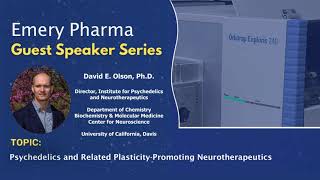
David E. Olson describes psychoplastogens, molecules that regrow brain connections after a single dose. Ketamine and classic psychedelics like psilocybin restore synaptic function in depression and PTSD models by activating the 5-HT2A receptor. Olson's lab is developing non-hallucinogenic versions like tabernanthalog that keep the therapeutic effects without the head-twitch response.
Chapters
Hi, my name is Ron Najafi, I'm the CEO of Emory Pharma. It's an honor to have Professor Olson here today virtually. Professor David E. Olson studied chemistry and neurosciences at Stanford University and the Stanley center for Psychiatric Research at the Broad Institute of MIT and Harvard. His academic lab at UC Davis discovered that psychedelic promotes structural and functional neuroplasticity in the cortex. They coined the term Psychoplastogen to describe small molecules that produce rapid and long lasting psychedelic and ketamine like effect on neuronal structure after a single dose. And they invented the non hallucinogenic Cycloplastogen capable of producing sustained therapeutic effect in preclinical model after a single dose. Professor Olson's expertise spanned central nervous system medicinal chemistry, molecular cellular neurobiology and behavioral neuropharmacology. He he's an Associate editor at the ACS Chemical Neuroscience and has received numerous awards including the Jordi Folch PI Award from the American Society of Neurochemistry, the Sigma Chi Jung Investigator Award, the Life Young Investigator award, Sacramento business journals, 40 under 40among many others. He's the founding director of the UC Davis Institute for Psychedelic and Neurotherapeutic and is the co founder and the Chief Innovative Innovation Officer of Dellix Pharma Therapeutics. In 2019, myself personally and my family, we lost our father to Alzheimer and dementia. It was then that we realized how little medical intervention was we had at our disposal. It was like watching my father slip away out of our hands. That's when my family established the nejafe Award at UC Davis Department of Chemistry. The faculty and postdoc at UC Davis have a chance to compete for a $30,000 annual grant. The research application needs to be focused on sparking innovation at the interface of chemistry, biology and NEUR with the hope of developing drugs for CNS diseases. Our first year recipient for the award, which was back in 2020, was Professor David Olson. So it is our pleasure to host David. Today I had a chance to review his slides and I can assure you there are lots of gold nuggets within his presentation. So stay tuned. Here's Dr. Olson.
Thanks Ron. And before I start, I just want to thank you and your family again for your generous support of our research. I'm very hopeful that some of the discoveries that we've made will eventually make it into human trials and hopefully impact the lives of patients. Before we begin today, I need to tell you that in addition to my academic appointments at UC Davis, I'm also a co founder and the chief innovation officer of Delics Therapeutics. But all the work I'm going to tell you about today is worked on in my academic laboratory. So our group is really interested in finding new treatments for a variety of brain disorders. But today I'm going to focus on the so called stress related neuropsychiatric diseases. These are conditions like depression, post traumatic stress disorder, and substance use disorder, Conditions that are precipitated or exacerbated by chronic stress. Now, a hallmark of all of these illnesses is physical changes in the structure of the brain, particularly the atrophy of key neurons in the prefrontal cortex. Now, that's perhaps unsurprising, because the prefrontal cortex projects to a whole bunch of subcortical regions that regulate things like mood, motivation, fear, and reward. Now, our current therapeutic strategies for treating these diseases, unfortunately, are far from perfect. They have largely relied on trying to rectify chemical imbalances in the brain through the use of things like selective serotonin reuptake inhibitors. But unfortunately, these medicines take weeks to months to demonstrate any kind of efficacy. And about a third of all patients will never respond to these traditional types of medicines. Our group has been trying to pioneer a new approach, one that involves the use of small molecules to selectively modify specific circuits in the brain to produce long lasting behavioral changes. And we reasoned that perhaps one of the best ways to impact the neural circuit was to physically change the structure of neurons within that circuit. And so, for the purposes of today's talk, I'm gonna be focusing on the structure of these layer 5 pyramidal neurons in the cortex that tend to atrophy in response to chronic stress. So we started studying this class of compounds that we call psychoplastogens. These are just chemical stimuli that induce structural neuroplasticity, such as the growth of these new dendrites. And if you zoom in on one of these dendrites, you'll see the growth of these little tiny nubs called dendritic spines. And dendritic spines are the sites of excitatory synapses in the brain. So these changes in structural plasticity often manifest as full functional changes that you can measure with traditional techniques like electrophysiology. Now, one of the best known psychoplastogens is this molecule on the left here, ketamine. Ketamine is a dissociative anesthetic that's been used in medicine for many, many years. But more recently, it was approved by the FDA for treatment resistant depression. And the way that we believe ketamine is working is by promoting the growth of, of those really critical cortical neurons. So here I'm just showing you some cultured neurons that were treated with ketamine. And you can see they have more of these dendritic branches. And then on the bottom here, these green dots are simply representing synapses. So you can see that ketamine promotes both neuridogenesis as well as synaptogenesis. Now, I'm showing you some work from the late Ron Duman's lab and George Akajanian. And what they were able to demonstrate is that chronic stress leads to that cortical atrophy, the loss of those dendritic spines that I was telling you about, which manifests as synaptic deficits. But what is really remarkable is that a single dose of ketamine can rapidly regrow those lost dendritic spines and reestablish synaptic connectivity in the cortex. So I think that ketamine is super exciting for neuropsychiatry because it's pushing us in a completely new direction, a direction that involves the physical healing of the brain and fixing these damaged neural circuits rather than simply trying to treat disease symptoms. However, ketamine is not a perfect drug. It lacks what I would call circuit level selectivity. One of its primary targets is the NMDA receptor, which is found broadly across the entire brain. And so it has some non selective effects that are less than ideal. So when we got into this space and we were trying to identify molecules that had ketamine like effects on neuronal structure, we were trying to think of compounds that would hit targets that are selectively expressed exactly in the regions of the brain that we care the most about. And that led us to study the serotonin or 5ht2a receptor. The 5ht2a receptor is heavily expressed on these layer 5 pyramidal neurons in the cortex that tend to atrophy in response to chronic stress. You can see in the rodent brain, it has high expression in the anterior parts of the brain, with less expression as you move towards the back of the brain, and very little expression in structures like the hippocampus or the striatum. Now, if you're interested in trying to modulate the 5ht2a receptor, one of the first classes of drugs that you think about are the classic serotonergic psychedelics, molecules like LSD and psilocin, which comes from magic mushrooms. So one of the very first experiments that we did is we simply cultured cortical neurons, treated them with Psychedelics and looked at their morphology. And as you can see here, compared to the vehicle treated neurons in the middle, the neurons treated with the psychedelics, DMT and LSD have substantially more complex dendritic arbors. And if you do some super resolution microscopy and zoom in on one of those dendrites, you'll also see the growth of these dendritic spines and these filipodia like structures. Now, there's a number of ways that we can quantify structural plasticity in the brain, but I want to focus on one of them because you're going to see a whole bunch of these graphs throughout the talk. We do something called shoal analysis. It's a pretty simple technique. All we do is we draw these concentric circles of increasing radii starting at the soma of the cell, and we simply count the number of times one of these branches crosses one of these circles. And so you can produce these really beautiful shoal plots like the one on the right, where on the x axis is the radii of those circles and on the Y axis is the number of crossings. But typically the way we like to represent the data is simply looking at the maximum number of crossings. So for something like the vehicle, that would be about 7, and for something like DMT, that would be about 11. So looking at these NMAX plots, we can see that psychedelics from three distinct chemical classes. Ergolines like lsd, tryptamines like DMT and amphetamines like doi, they're all really good at increasing the complexity of dendritic arbors. Now, for all of these experiments that we were doing at this point, we were treating the cells for three days and then looking at structure, we know that in vivo that's not what's going on. In vivo, you give a psychedelic, it rapidly gets into the brain and then it's rapidly cleared. But they seem to produce these very long lasting effects. We were wondering what is the shortest stimulation period that we could get away with and still produce long lasting changes in neuronal structure? Instead of stimulating for three days, we just stimulated for 24 hours or an hour, or just 15 minutes. And then we take the drug away and give the neurons time to grow. And when we do that, we find that even this 15 minute stimulation is sufficient to produce a long lasting increase in dendritic growth. Now you have to give the neurons time to grow. If you stimulate for an hour, fix and stain the cells and image, you see no changes in cortical neuron structure. But if you give them time to grow, you'll see very robust changes. Now, in order to demonstrate this in vivo, we relied on a very rapidly cleared psychedelic, dimethyltryptamine. The half life of DMT is only about 10 minutes in a rodent. And so for this experiment, what we did is we gave a single IP injection of DMT, waited 24 hours, a time period when all of the drug has been cleared from the body. We then sacrifice the animal, take tissue and look at structure via Golgi staining and function via electrophysiology. We find that even after the drug has been cleared from the body, we see this very robust increase in dendritic spine density. And we also see changes in functional plasticity. In particular, we see an increase in both the amplitude and the frequency of these spontaneous excitatory postsynaptic currents. Now, ever since we published this work way back in 2018, it has been replicated and extended by a number of groups across the world. But I want to focus on one study in particular because I think it's quite remarkable. And this was a study done by Alex Kwan's group when he was at Yale. He has since moved to Cornell. And Alex was really interested to see just how long do these effects last for. He gave a single dose of psilocybin to male and female mice, and he found that even after a single dose, he saw increases in dendritic spine density that lasted for at least a month, which is quite remarkable. Now, in addition to their long lasting, sustained effects after a single dose, the other thing that's really unique about psychedelics is that they seem to have this trans diagnostic panacea like property. They work across a variety of indications. Depression, ptsd, substance use disorder. And I want to emphasize that these drugs are not panaceas. We think the reason they work across indications is because they're targeting a very critical hub in the brain, the prefrontal cortex. And we know from a whole bunch of opto and chemogenetic studies that there are circuits that originate in the prefrontal cortex that do things like regulate drug seeking, behavior, control, the expression of fear, memories, impact, motivation, et cetera. And so to summarize a lot of work very, very quickly, in general, what we find is that psychoplastogens produce long lasting behavioral effects in behaviors that are known to involve PFC circuitry, such as the enhancement of fear, extinction, learning, or producing antidepressant like effects in something like the force swim test. Now, at this point, we were very interested in trying to understand the mechanism by which these drugs produce their effects on cortical neuron structure. And so we started with some selective antagonist studies and we found that if we used a antagonist of the 5ht2a receptor, we were able to block the effects. The 5ht2a receptor is known to mediate the hallucinogenic effects of psychedelics. And so this is one of the first receptors we looked at. We also wanted to probe a couple of downstream kinases that had been implicated in the mechanism of ketamine. One is track B, which is the high affinity receptor for brain derived neurotrophic factor, and the other is mtor, which is necessary for the production of all of the proteins that are required for a neuron to change its morphology and to change its synaptic weights, all those structural proteins and those ion channels. Previous work had shown that if you block mtor, track B, you block the effects of ketamine. And we see something very similar here with psychedelics. Now, at this point, we realized that the antagonist that we were using to block the 2A receptor was not perfectly selective. It hits a couple of other receptors. So to be really, really certain that it was the 2A receptor mediating these effects, we relied on genetic knockout models. So here I'm showing you. In wild type animals, when you treat them with the psychedelic 5 methoxy DMT, you see this really nice increase in dendritic spine density in the pfc. But in the knockout animals, that effect is completely gone. You see very similar things in terms of functional plasticity. Again, wild type animals, you get a robust effect in functional plasticity, but in the knockout animals, the effects are gone. So to determine the role of the 2a receptor in behavior, we ran an experiment relevant to depression. And this is a particular type of the forced swim test that is a little more challenging, I would say. Traditional antidepressants tend to not work in this, but these next generation psychoplastogens tend to be more effective. So on the first day, we run a force swim test to kind of stress the animals out. And you can see that all the groups at this point are exactly the same. We then give a dose of 5 Meo DMT and we do a behavioral assay just to make sure that the dose is getting into the brain and doing what we expect. But then, 24 hours later, after the drug has been cleared from the body, we assess the antidepressant potential. And you can see that in the absence of the 5 HT2 antagonist, Catanserin, 5 MeO DMT causes this robust antidepressant. Response. But if you pre treat with the antagonist, you can completely block that. Now, in addition to looking at for swim test behavior, which is relevant to, I would say, motivated behaviors, we also wanted to assess anhedonia. And the way we do this in mice is using a sucrose preference test. So mice much like myself will prefer a sugary beverage over just plain old water. And you can measure this preference. And you can see that in wild type and the 5ht2a knockout animals, you have a really robust preference for the sugary sucrose containing solution. But you can induce an anhedonia like phenotype by giving chronic stress. And in this case we're giving chronic corticosterone injections. And then we can do another sucrose preference test on day 12 and you can see this big reduction in their sucrose preference. Now what is really interesting is that on day 13 we give a single dose of psilocybin, come back on day 14 and look for sucrose preference. And we can see that in the wild type animals we see a complete rescue of the sucrose preference. But that effect is completely gone in the 5ht 2a knockout animals. So at this point we're feeling pretty confident that the 5ht2a receptor is mediating the psychoplastogenic effects of the compounds and at least some of their antidepressant behavioral responses. But there was a piece of data that really bothered us and that was the fact that this molecule here on the left, serotonin, could not promote cortical neuron growth. That had us scratching our heads for a long time, because serotonin is a very good agonist of the serotonin 2A receptor. This didn't really make a whole lot of sense until we did this structure activity relationship study. Basically we were making small structural changes to serotonin like molecules. And we found that as we started to add methyl groups, going from one methyl group to two methyl groups, we saw an increase in the ability of these molecules to promote cortical neuron growth. And if you look at the shoal plots, it's pretty pretty striking. Going from tryptamine, you see no difference compared to vehicle N, methyltryptamine you see a little bit. But dmt, dimethyltryptamine, you see a very, very big bump. Then we tried to correlate activation of the 5ht2a receptor with efficacy in this neuroplasticity assay and we didn't see any efficacy. If anything, we saw maybe a negative correlation, but Then we correlated the lipophilicity of the drugs and we saw a really nice positive correlation with with their ability to promote cortical neuron growth. And this led us to the hypothesis that maybe the reason that serotonin cannot promote cortical neuron growth is because maybe it can't access the target. Maybe this particular G protein coupled receptor is not existing on the cell surface. Maybe it's existing on the inside of the cells. And serotonin is simply too polar to cross the nonpolar plasma membrane and access that receptor population. But the psychedelics, which are very lipophilic, can easily cross the membranes. So to start to address this question, the very first thing we did is simply look at where the receptor is localized. So for this experiment, we simply expressed the 5ht2a receptor and the beta2adrenergic receptor in hex cells. They're tagged with CFP. So we use CFP as a control. You can see that CFP is expressed everywhere in the cells, but the GPCRs are expressed pretty much where you would expect them to be. There's a little bit intracellular, but there's a lot on the outside of the cells. And here's just a membrane marker that indicates the boundaries of the cells. But when you express these two GPCRs in cortical neurons, you see something very different. You see that for the five HT2A receptors, you primarily see an intracellular localization of the receptors, whereas the beta 2 adrenergic receptor is still expressed on the cell membrane quite a lot. We did a whole bunch of follow up studies to see what sub cellular organelles that the 5ht2a receptor might be co localizing with. And what we found is that when we use this marker of the Golgi complex, we saw a really nice overlap with five HT2A receptors in these cortical neurons. But when you look in hex cells, you don't see as good of an overlap. You can see here, there's lots of 5 HD2A receptors that don't overlap with the Golgi, for instance. So we think that in neurons, cortical neurons, the 5ht2a receptor is existing primarily inside the cell and primarily at the Golgi. Now, I should emphasize we're not the first to discover this localization. This has been known for many, many, many years. But no one really appreciated what the functional consequence of that intracellular population was. To probe that, we first developed a few chemical tools. We started by just taking these Membrane permeable compounds in green, two agonists here and an antagonist here. And we just converted them into membrane impermeable versions that are permanently charged. So we just methylated DMT to make trimethyltryptamine, put a phosphate group on this hydroxyl to make psilocybin, and methylated this nitrogen to make methylated catanserin. And then we simply run the assays under two sets of conditions. The first set is just normal neurons. The second set is under electroporation conditions. What we do there is we transiently apply a voltage. And what that does is it opens pores in the membrane that allows the impermeable compounds to cross the membrane. The pores then close and then the impermeable compounds get stuck on the inside of the cells. When we run the assay without electroporation, we get exactly what you would expect. The permeable compounds agonist, DMT and psilocin can promote cortical neuron growth. The impermeable compounds TMT and psilocybin cannot. Similarly, the membrane permeable antagonist can block DMT induced plasticity. The impermeable antagonist can cannot. But when we electroporate the cells, we see something very, very different. In this case, we can see the impermeable agonist TNT and psilocybin. They can promote neuronal growth and the impermeable antagonist can block cortical neuron growth. That really suggested that the target is on the inside of the cell. Now, this is a neritogenesis assay. We did the same thing for a spinogenesis assay. And again we see the permeable compounds in green promote growth, the impermeable compounds do not. Now, at this point, we reasoned that if it's all about the location of the receptor, serotonin should be able to promote growth if we could just get it to the target. And so for this set of experiments, we relied on sparsely transfecting cortical neurons with a transporter of serotonin, this protein called sirt. So I love this experimental design because within the same culture you can compare the two different genotypes. So I'm showing you here two neurons side by side. The one on the left here expresses the transporter, the other one doesn't. Similarly, these two neurons here, the one on the right expresses the transport and the one on the left doesn't. And so what we see is that when you treat neurons of either genotype, again in the same culture with vehicle, you see no change when you treat them with dmt, you see increased growth in both genotypes, which makes sense because DMT can just passively cross the cell membrane. But when you treat them with serotonin, you only see growth in the neurons capable of importing serotonin. Now, to follow up on this experiment in a little more detail, we ran this study in the presence of a 5ht 2a antagonist and a blocker of the serotonin transporter. So what I'm showing you here in this bar graph, this is essentially a replication of the last slide that I just showed you. Serotonin can promote growth, insert positive neurons only. But then if you treat with catanserin, you can block this effect, suggesting it's 5ht2amediated. And you can also block the effect with citalopram, suggesting that serotonin needs to enter through the transporter in order to turn on the pathway. But when you do this with DMT instead of serotonin, now you see again, a replication of that last study. It works in both genotypes. You see that catanserin blocks the effects, again because it requires 2A. But citalopram no longer can block the effects of DMT because DMT does not require the transporter to access the 2A receptor. Now, those were neuridogenesis experiments. We did the same thing with spinogenesis experiments. And so again, here I'm showing you that DMT promotes spinogenesis in both genotypes, but serotonin can only promote spine growth in cells capable of importing serotonin. Now, to demonstrate this in vivo, we relied on using a virus to express the serotonin transporter on those cortical neurons. And so normally they don't have the transporter, so they can't import serotonin. But after three weeks, to allow for viral expression, we then give a single dose of this compound, parachloral amphetamine. It's a serotonin releasing agent. It causes presynaptic release of serotonin. And then we simply DO spine analysis. 24 hours later, what we find is that in the neurons that have the transporter, we see a really nice increase in spinogenesis compared to the neurons that lack the transporter. Now, if the spine growth is responsible for the antidepressant effects, we would expect to see something similar. We ran a very similar experiment, except for after expression of the transporter, we ran a novelty induced locomotion assay just to show there's no difference between the genotypes, and there wasn't. We then run A for a swim test, again to show no difference between the genotypes. We then give parachloroamphetamine, come back 24 hours later and measure antidepressant like effects. And what we see is we get an antidepressant response only in the animals that have the ability to import serotonin into those cortical neurons. Okay, so at this point, we're feeling pretty confident that the intracellular population of 5ht2a receptors is really critical for psychedelic induced neuroplasticity and the antidepressant effects. However, there, there's a problem, and the problem is that the 5ht2a receptor also mediates the hallucinogenic effects of psychedelics. In While I definitely think these first generation hallucinogenic medicines are going to help some people, their clinical scalability is limited. These compounds, in order to be administered safely, have to be given under the supervision of a healthcare provider, which drastically increases the cost and the complexity of the treatments. And so we were wondering if it might be possible to decouple the hallucinogenic effects of the drugs from their effects on neuroplasticity and their beneficial effects on behavior. And we thought it might be possible because there were known agonists of the 5ht2a receptor that are non hallucinogenic. This receptor exhibits a high degree of functional selectivity. And so in order to try to identify these non hallucinogenic cycloplastogens, we needed an assay that was very good at predicting hallucinogenic potential. And so we turned to this mouse assay called the head twitch response. You can see the behavior there on the left. It's this rapid rotational movement of the head, and it's very characteristic of serotonergic psychedelics. And so what Adam Halberstad's group was able to show is that across over 30 structurally distinct psychedelic compounds, there's a really beautiful correlation between human hallucinogenic potency and potency in this head twitch response assay. And so I'll give you an example of kind of just how small the structural change can be in order to completely change the hallucinogenic properties of a molecule. So we started with this compound, dmt, a very potent hallucinogen, and we converted it into this ISO DMT analog you can see here in terms of its psychoplastogenicity compared to ketamine as the positive control, the compounds are essentially the same. They have, you know, equipo, but their hallucinogenic potentials are very different. So I'm showing you a couple examples here. 5 Meo DMT in blue and 6 Meo ISO DMT in orange. You know, if you had a space filling model of these two compounds and you're holding them in your hand, you have a really hard time telling them apart. The only difference really is that this 5 Meo DMT has the ability to engage in a hydrogen bond donor event which is gone in the, in the ISO DMT counterpart. And even though they have comparable effects on neuroplasticity, very different effects in the head twitch assay. 5 Meo DMT causes this nice dose dependent increase in head twitch response. You don't see that with the 6 Meo ISO DMT analog. Now the head to response is great because it's highly predictive, but it's not very useful for drug discovery, mainly because the throughput is quite low. And so we realized that we needed to develop a cellular assay for hallucinogenic potential. And the way we did this was actually quite simple. We took the serotonin 2A receptor, which again is known to mediate the hallucinogenic effects of psychedelics, and we simply lopped off the third intracellular loop of this GPCR and we fused on a circular permuted green fluorescent protein. Now the way these CPGFPs work is there's basically a cut in the beta barrel that allows it to be open, that enables water to get in, quench the fluorescence of the fluorophore. But when a ligand binds to the receptor, it can induce a conformational change that closes the beta barrel, protects the fluorophore and allows fluorescence to turn on. So you can see that this agonist turns on the sensor really, really nicely. But these two antagonist compounds either do nothing or they actually turn it off slightly. And so we got very excited when we found that potency in this biosensor assay correlated really nicely with human hallucinogenic potency. So we started calling the biosensor cyclite. Now, one of the first experiments that we did with cyclite is we used a virus to express it in the prefrontal cortex. We then gave a single administration of 5 methoxy DMT. You can see that very rapidly cyclate fluorescence turns on. But what's interesting is it turns on at the same time that the head twitch behavioral response turns on. But what was surprising to us is that the head twitch behavioral response wanes over time where a cyclite fluorescence remains elevated. Now, if you give an injection of the vehicle or you give an injection of the antagonist, you see no change and a decrease in fluorescence, respectively. Now, I would say that probably the most exciting day came when we saw these data and we were comparing hallucinogenic compounds in green with their non hallucinogenic congeners in purple. And you can see that the difference between LSD and lyceride is subtle. Lisa Ride simply has a nitrogen inserted in here and that stereocenter kind of switched. And that's it in 5 Meo and 6 Meo DMT. The only difference is you move the Methoxy Group 1 position over on the indoor ring and that's it. But what is remarkable is that the hallucinogenic compounds turn on the biosensor, the non hallucinogenic compounds do not. Now, you might be asking yourself, maybe the non hallucinogenic compounds are not binding to the sensor. But to address that question, we can simply run the assay in antagonist mode. We can give a little bit of agonists to turn on the biosensor and then titrate in increasing quantities of the non hallucinogenic ligand. And you can watch the fluorescence intensity decrease. And that's what you see here on the right. So to facilitate our drug discovery efforts, we simply made a stable cell line that expresses cyclate. We call these sili2 cells. These cilia 2 cells are really nice because they exhibit some baseline fluorescence. So inverse agonists turn off the fluorescence and agonists turn on the fluorescence. And we tested a whole bunch of compounds in this assay I'm showing you. On the x axis is agonist mode. On the Y axis is antagonist mode. And this black dot right here, that's the vehicle control compounds that increase cyclite fluorescence more than one standard deviation from the vehicle. These are known hallucinogenic compounds in red. Compounds that decrease cyclate fluorescence more than one standard deviation in antagonist mode. These are known non hallucinogenic compounds in blue. And we can simply mathematically combine agonist and antagonist mode data to produce this ligand score. On the left here, you can see that compounds with a red ligand score are predicted to be hallucinogenic. Compounds with a black ligand score are predicted to just not bind to the sensor period. And compounds in blue are predicted to be non hallucinogenic ligands. What's really interesting about this class is you'll see traditional antagonists like the MDL compound, which is also known as volumanserin, catanserin and antipsychotics like haloperidol, but you'll also see these non hallucinogenic agonists like glyceride. Now, I want to spend just a little bit of time telling you about these two compounds. I'll start with 5 Bromo DMT. What was really interesting about 5 Bromo DMT was that we had synthesized the entire halogenated series, going from fluoro to chloro to bromo, just increasing the van der Waals radii of those atoms. And the biosensor predicted the fluoro and the chloro compound to be hallucinogenic and predicted the bromo compound to be non hallucinogenic. And that's exactly what we see in the behavioral assays. The fluorocomb produces robust head twitches at all doses. The chlorocompound produced this nice dose dependent increase, but the bromo compound did not produce head switches at any dose tested. Now we also found this compound that we call az, named after the student who synthesized it. Kind of looks a little like 5 Meo DMT. The biosensor was predicting that it was going to be non hallucinogenic and it didn't produce a head twitch up to even these massive 100mg per kig doses. Now, a therapeutic dose of AZ is a lot closer to 10mg per kg. Now when we tested this in assays relevant to depression head to head with ketamine, we found something really interesting. We found the AZ had comparable potency to ketamine in vitro for inducing cortical neuron growth. We also found that it had rapid after 30 minutes and sustained lasting at least a week in the four swim test that was comparable to ketamine. And then finally, we use this genetic model of depression developed by Bill Wetzel's group at Duke. These VMAT2 heterozygous mice. These mice just have some baseline depressive phenotypes. And here I'm showing you that sucrose preference test again. And you can see that the wild type mice have a very robust sucrose preference, but the heterozygous mice do not. A single dose of AZ can rapidly rescue this anhedonia. And what's really remarkable is that the effect lasts for quite a long time. It lasts for at least a couple of weeks before the genotypes start to differentiate from each other. Okay, so at this point, we have been playing these kind of medicinal chemistry tricks on compounds from every single major psychedelic clinic class. Tryptamines like psilocybin, amphetamines like, you know, doi, mdma, Ergolines, like lsd. But I want to spend a little bit of time to tell you about our work on this compound right here, ibogaine. Now, ibogaine is not a traditional classic psychedelic, you might call it an atypical psychedelic. But ibogaine has some pretty remarkable properties, at least as anecdotal reports and some open label clinical trials would suggest. Those studies have suggested that a single dose of ibogaine can have anti addictive properties that last for at least six months. And if you take a second dose, they can last up to three years. And that is just really remarkable and really indicates plasticity mechanisms to me. And so you might be asking yourself, well, if ibogaine is so efficacious, why are we not using it to treat the opioid epidemic in this country? And the answer is, ibogaine is a terrible drug for a variety of reasons. Number one, it's hard to come by. There are no de novo total synthesis that can produce ibogaine enantioselectively in the quantities required for clinical trials. And getting it from the natural sources has a whole bunch of problems associated with that. Number two, ibogaine is incredibly greasy and it's very insoluble in water, which makes formulation quite challenging. Of course it has these hallucinogenic properties. But the worst thing about ibogaine is that it binds to her channels in the heart and causes cardiac arrhythmias. Several people have actually died during ibogaine treatment for their opioid use disorders. Our approach to this problem was to use something called function oriented synthesis. This is a really simple concept popularized by Paul Wender at Stanford. And, and the idea is, can you take a complex structure like ibogaine and distill it down into the features that are essential for the properties that you care about? And if you look at the structure of ibogaine, it really has kind of three distinct structural motifs. It's got this indole group, it's got this seven membered tetrahydro azepine ring and it's got this bicyclic isoquinuclide. So all we did was to simply start deleting bonds. If you delete the bond between the indole and the isoquinucleidene, you arrive at compounds that look like scaffold one. If you delete the isoquinuclein altogether, you arrive at compounds that look like Scaffold 2. I'm not going to take you through all of the medicinal chemistry that we did. I'M just going to show you some of the high level results. And basically what we found is, was that compounds that retained the ASAP and lacked the isoquinucleidine, well, they were pretty good for the most part at promoting cortical neuron growth. But the opposite wasn't true. Compounds that contained the isoquinucleidine but lacked the azepine, they were pretty bad for the most part. So we decided to prioritize this compound here for further study. We made one more small structural tweak to it. We moved the Methoxy Group 1 position over to produce this compound. The compound on the left looks just like ibogaine, except it lacks the isoquinucleidine. So we started calling it ibogaine a log or ibg. But the compound on the right looks a lot like the natural product tabernanthine. So we started calling this tabernanthalog or tbg. Now, unlike ibogaine, TBG can be synthesized in just a single step in gram quantities using this Fischer indole cyclization. And it has a lot of really desirable properties. First and foremost, it's completely soluble in water, which makes handling it really easy. It is still a potent agonist of the 5ht2a receptor and has some other unique properties. And so here is one example that I'm going to show. I want to just emphasize this particular structure activity relationship study because I think it's really quite remarkable. So 5 methoxy dimethyltryptamine is a substructure within ibogaine and it's a very potent hallucinogenic compound. And you can see that 5 Meo DMT produces a very robust head twitch response. But if you simply confirmationally restrict the dimethylamino group to the two position of the indole to make ibg, you can see that you drastically lower the hallucinogenic potential. And if you move the Methoxy Group 1 position over to make TBG, now you've completely eliminated the hallucinogenic potential. So what about the cardiotoxicity? So the other thing that was really interesting about TBG is that by cutting away all that extra grease in the isoquinucleidine, we drastically reduced the potency of TBG for Herg. So Ibogaine's potency at Herg is about 1 micromolar. You can see that TBG is 150 fold less potent. And you can actually visualize this using these immobilized zebrafish here on the right. So you can see the heartbeat of a vehicle treated fish you can see the slowing or the bradycardia induced by ibogaine, but you just don't see that with TBG treated fish. Now one way to measure the propensity of a drug to induce a cardiac arrhythmia is simply to take a ratio of the atrium to ventricle beats per minute. Here we see that ibogaine is very good at causing cardiac arrhythmias, as is the positive control sir indole. But TBG looks just like the vehicle control. Now in addition to the cardiotoxicity, TBG has some just general toxicity issues associated with it. And you can look at these, these pie charts, but I think a picture is worth a thousand words. This is what a vehicle treated fish looks like. This is what an ibogaine treated fish looks like. They look very different, but a TBG treated fish looks just like the vehicle control. Now it's great that we produced a safer version of ibogaine, but none of this matters if it doesn't have ibogaine like efficacy. So the first thing we did is verify in vitro that TBG is a potent psychoplastogen. But this is an in vivo study looking at that same effect. And the way this works is we have these mice that are genetically modified to have their neurons in the cortex labeled with a fluorescent protein proteins. So we can visualize them. We simply cut a window in the skull and we use a two photon microscope to look at a section of dendrite. We then give a single dose of TBG, come back 24 hours later and look at the exact same section of dendrite. And that allows us to quantify the number of spines that were formed, like this one in blue, and the number of spines that were eliminated, like this one in red. And we find that TBG does not really impact spine elimination, but it does seem to increase spine formation and it does so to a comparable extent as the hallucinogenic drug doi. Now again, if the spinogenic effects of the compound are really important for the therapeutic effects, we would expect to see kind of broad behavioral changes. And that's exactly what we see. We see that TBG has antidepressant like effects in the four swim tests comparable to ketamine. We also see a very robust anti addictive response in this two bottle choice paradigm relevant to treating alcohol use disorder. And then finally we used a model of heroin use disorder and we gave TBG at three different time points during self administration of heroin, during the early stages of extinction or withdrawal, and then immediately prior to Cued reinstatement, it didn't really matter when we gave the drug. We saw this long lasting protective effect on heroin seeking behavior. Now, finally, I just want to emphasize that TBG has the potential to actually heal damaged neural circuits. What I'm showing you here is some calcium imaging from the somatosensory cortex of mice. Now, mice are very tactile creatures. They like to explore their environment with their whiskers. And every time they whisk, you see this really nice coordinated calcium response in the somatosensory cortex. However, unpredictable mild stress can completely disrupt this coordinated response. But what is really remarkable is that a single administration of tbg, or long after the drug has been cleared from the body, can completely restore this coordinated response, suggesting that it has healed this damaged neural circuit. So to summarize, what I tried to tell you about today is that in the healthy brain, in your cortical neurons, there's lots of dendritic branches and lots of these dendritic spines. But in many brain disorders, including many neuropsychiatric diseases, and in many neurodegenerative diseases, you see a physical atrophying of the brain, a loss of these dendrites, and a loss of these dendritic spines. But psychoplastogens, be they hallucinogenic compounds like psychedelics or non hallucinogenic compounds like tbg, are very good at regrowing these atrophied dendritic arbors. So finally, to summarize, I told you today that psychedelics are very good at promoting structural neuroplasticity through the activation of this intracellular population of 5 HT2A receptors. I told you that cyclate is very good at predicting hallucinogenic potential and so has applications in drug discovery. I told you that TBG and AAZ are potentially safer cycloplastogens. I just like to finish by thanking these key collaborators that played important roles in this work. In particular, I think Lynn Tiana was a really great collaboration working with her group on the development of cyclite. And I'd like to thank these funding agencies on the right. In particular, I'd like to thank the Najafi family for, for their generous gift. Unfortunately, we have not published the work that we did as part of the Najafi award yet. But I want to tell you right now, compared to everything else that I told you about today, that work is the most exciting. We have a really phenomenal compound. It's currently in revision. And so I'm hoping that maybe in a few, few months, maybe I can come back to Emory in person and give a lecture on that work. Really, really exciting stuff. We're hopeful that it's going to have some real impact on neurodegenerative diseases. And finally, I just want to thank all of the really incredible students that I get to work with every day. Very creative, talented, hard working bunch. I've highlighted the names of just a few people whose research I focused on today. And with that, I just want to thank you for your attention. I'd be happy to answer any questions.
More from Dr.

Hippocrates said, let thy food be thy medicine #obesity #metabolicsyndrome #lustig #shorts #health



Bill Wilson, MD: Why GLP-1 Drugs Are Overpriced—and How Compounding and Competition Could Fix It
Nitrosamine in Pharmaceutical Products? A Deep Dive into Risk Management by Ron Najafi, Ph.D.

The Subcellular Processes That Belie All Chronic Disease. The Hateful (or Grateful) Eight #lustig

Bill Wilson, MD: GLPs May Reverse Carb Syndrome—Helping Patients Reach Zero and Sustain with Diet

Why Everything You Knew About Obesity Is Wrong | Dr. Robert Lustig on Sugar & Metabolic Syndrome
Robert Lustig, MD: How Fructose Fuels Fatty Liver, Oxidative Stress, and Metabolic Disease #fructose
Robert Lustig, MD: The 3 Types of Fat Fueling Insulin Resistance and Disease #insulin resistance

Bill Wilson, MD: GLPs May Reverse CARB Syndrome. Helping Patients Reach Zero and Sustain with Diet