Cardiovascular Examination p1
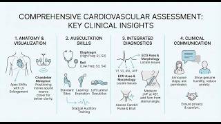
Dr. Hassan Bencheqroun teaches cardiovascular examination using surface anatomy landmarks, the APTM auscultation points, and maneuvers like left lateral decubitus to detect heart sounds and murmurs. He covers JVP measurement, apical impulse palpation, and how ECG leads map to coronary territories for infarct localization.
Chapters
Yes. So when we're talking about cardiovascular exam, the surface anatomy matters because it is like giving you a map. It's like your map, not just for the, um, the auscultation, but also your EKG leads. And we're going to have a session only for EKG, how to place them, how to interpret them. And I would like to decomplicate it. Okay, when you look at someone's chest, I want you to feel like you have Superman x-ray vision. What I mean by that is when you see them, you see through them. Let's say, for example, you see someone and that person has been stabbed. If they've been stabbed in the chest, it depends where that's going to land. If they've been stabbed in the chest, and this is unfortunate, but it's not, it's, it's not like unlikely for you to see it because you're going to be working in the emergency room or Or even something happened. For example, this guy. This guy has been stabbed in the chest. You can see that the blade is going behind the sternum, right in the middle. So the next question is, What chamber of the heart is lying behind the sternum? Who can tell me that? I am here when we are comfortable. Though you are uncomfortable, that's where it sticks in your mind. We complain that we go into traditional classes and the teacher just wags the finger at us, just I did not forget to be compassionate. And I asked, I was going through, here's what I'm going to do, here's what I'm going to do. And there were days where it, no matter what, it just made me uncomfortable. Guess what? That uncomfortable feeling loses power over you once you say it. Forgive me, Mrs. Smith, I am a little bit nervous. This is my first time in a long time I'm examining a patient. I was taking my boards. Until now, and I just got back to the bedside. So if I do anything that's off, please understand that it's probably me being uncomfortable and feel free to correct me. Does this make sense? You appeal to their caregiving sense and you show them innocence, and that goes very smoothly by then. Okay. Then when we talk about the chest, the chest has a system. Whenever you are in that system, what are the bars in that system? The blue are the ribs. The yellow is costal cartilage. And then you have the sternum. And the sternum is like a tower, but leaning. The top of the tower, the manubrium of the sternum right here, is actually leaning forward. So we start to call these things by names. We have the sternal angle. We have the midclavicular line. This is the clavicle. The midclavicular line comes through here. You have the 5th intercostal space. Okay, so this is how we speak to where's that landmark. You say, on the intersection of the mid-clavicular line or the mid-axillary line if it is off to the side underneath the armpit, mid-clavicular line on the right at the 5th intercostal space, proximal to the sternum. That's how we speak. Does this make sense, you guys? Excellent. And we talked about this. This is not a pyramid. This is a tilted, twisted pyramid. The right ventricle sits most anteriorly and directly against the sternum. The left ventricle forms the apex and projects left and downward. The atria are positioned at the base of the pyramid, and they are resting against the spine. So if you have a stab wound by a long thing in the back, it hits the atria. And then the tilt explains why the apex is found left of the midline. What is an APTM? That is where you're going to put your auscultation points. The A is aortic. The aortic valve can be seen in the right second intercostal space, which means it's going to be to the right of the sternum, proximal to the sternum at the right second intercostal space. Why do we have these complicated? Because they are precise. And if there is a blind— if there is a blind physician and you're speaking to them, they can visualize just by your words the precise location where you're pointing to. P is the pulmonary. It's the left second intercostal space across from the other one. Then you have T is the tricuspid. It's the left fourth and fifth intercostal space. And then the M is mitral. It's the fifth intercostal space at the apex. Okay? First, we do a palpation. You can start by the patient laying down. If you think about the heart as a chandelier hoisted from the neck and dangling inside the chest, if you put the patient on their back, then the heart will go backwards farther. But it's not too far. It's not like it's taking a 2-mile trip. It's just there, but it's slightly— so you go into the mid-clavicular line. Here it is. And you start to look for the apical beats. Now, this guy does it with their fingers as if he's taking the pulse. I will tell you, I don't do it that way. I do it with the palm of my hand, and I put the entire palm of my hand just like this because that is a surface area of my hand. And then it— I'm gonna feel it somewhere. And when I feel it, then I move it so that it hits here. I kind of move my hand, and when I move it and I finally hit it, then I see how far is it. Is it present? Is it displaced laterally and inferiorly? And is it a normal tap or is it a forceful heave? When you auscultate, you auscultate that lub-dub, lub-dub, lub-dub, lub-dub. But most people don't tell you that there's lub-dub. There is an actual gap between— there is a small gap, and you have to see that gap. It's not going to be— no, it's not one after the other. There is a gap. Okay, so there is an S1, S2, and a diastolic gap. You put the diaphragm or the bell. If you have a diaphragm that has— it's sitting right on top of the skin, so it's picking up the vibrations of the skin. When you flip it, the bell creates a sound chamber and it picks the low-sounding pitch sounds because they are echoed in that small little bell. So the diaphragm, you can hear the S1 and S2 and a lot of the murmurs. This bell, it's usually for S3 and S4. We talked about the maneuvers to amplify sound. Left lateral decubitus for mitral stenosis and S3. Leaning forward for aortic regurgitation. Respiration. Inspiration increases the right-sided sounds. Expiration increases the left-sided sounds. How does that sound? It sounds like something you can follow up with, correct? Now, there is When it comes to murmurs, I don't know about you. You're probably smarter, but I will tell you that when I was in your shoes, unfortunately, it just killed about killed me. I couldn't understand. People were getting it, and I just, I'm not hearing it. I don't know what you guys are hearing. I don't know. Where is this sound? How does this sound look? How does this sound feel? And that just bothered me. I felt like I was stupid, honestly. So what I learned is that's because we are doctors and we ask so much of ourselves. We want to learn it all at the same time. Don't do that. Learn One murmur at a time. This says one murmur a day. I did one murmur a week. And if I find a murmur, I go back every day and listen to it and do maneuvers and listen to it again, use another stethoscope, just so that I make it my project. It's going to be something I'm going to learn. So some people say, why am I going to do that? In order for me to when we have an echocardiogram that's gonna give me the diagnosis. It's not about, I know there is a murmur, so I'm going to diagnose it with a— I don't know that there is anything. I'm examining my patient for the first time. And if I don't know what murmurs are, I'm gonna hear something and I'm gonna think it's fine. But when I've heard a murmur before, I'm gonna hear and say, wait, that's a murmur. Let's call for an echocardiogram. Okay? Does this make sense, everybody? Yes? Thumbs up? Maybe? Questions? Very nice. Thank you. Alright. Now, there's not much that is left for us. I'm going to start you, but we're not going to do it today, the ECG mapping to the anatomy. What does that mean? It means that when we're talking about the ECG, every electrode has an axis going to it, an axis of electricity. And so wherever that electrode is, That's the tip of the arrow coming from the heart. Okay, so what that means is that the electricity going through that arrow from one place to the other, I'm going to look at how the QRS looks in there, and I'm going to see if there's anything abnormal. For example, if V1 and V2 are right here, the yellow and the turquoise blue. That means if V1 and V2 has a heart attack on them, the heart attack is in the septal area. Then I have to go back to the anatomy of the heart and say, what is the artery that feeds the septum? It's the left anterior descending artery, the LAD. So I'm looking at an EKG and I can already determine where, which artery might have the heart attack. Does this make sense, you guys? For example, if you find this right here, this is V1, but then you find in others like AVL, the left hand, AVF, the foot. If I have it in V1, AVL and AVF, that's where the heart attack is. Guess what? It's going to be in the right heart, and the right heart is fed by the right coronary artery. So the heart attack is in the territory of the right coronary artery. And that's how we use the EKG to map out where the problem is on the surface anatomy of the heart. Does this make sense? Fantastic. We are gonna have a session for ECG, but I wanted to get you excited about it. So in 60 seconds, how can you do a proper cardiovascular examination? Well, at first, you talk to your patient. And while you're talking to the patient, you look at the other vessels, not just the heart. You see at the bottom of their neck are the carotids. Doing this. Okay. So if the, if the carotids show If the carotids show that there is a If the carotids show that there is a Right there. So when you look at the carotid, what you saw was the auscultation was done by the bell. It does not get done with the diaphragm because you want to hear if there is a bruit, if you hear a whooshing sound. And what I mean by that is there might be a cholesterol plaque and the fluid going there becomes turbulent. But before you put the stethoscope, you also want to see how the carotid throbs. So we're going to take a look at his carotid before he puts the stethoscope on. Let's take a look. And it looks quiet. Sometimes it is not quiet. Sometimes the carotid is throbbing. And if it is throbbing, It means many, many things. It could be because there is an aortic regurgitation. So the aorta— aortic valve is open. And so you're sending a huge cardiac output because some of that regurgitates each time. And now the heart has to pump even more. And so there is a jet that comes out in the aorta and the jet goes to the carotids and you can see The carotid's constantly booming. That is important for you to make sure that you inspect before you auscultate. Then inspect the precordium. Is the precordium lifting off? And you inspect the jugular venous pressure. Are the jugulars completely engorged? If the jugular venous pressure is high, then that means there is some degree of heart failure. Okay. So let's see first how it looks like. We are going to show— this is how a jugular venous pulse and pressure is. Okay? Agreed? Yes? I'm waiting for reactions. Thank you, Muhammad. Okay, so usually the jugular venous pulse goes maybe halfway, about 5 centimeters, but here it goes all the way. So how do we measure that? Well, here's the internal jugular behind the sternocleidomastoid, right? So to measure the pulse, is where you lay the person on the head of the bed elevation at an angle. You turn their head to the left. You un-clear this area. You look for the sternocleidomastoid right here and the clavicular head. Those two give you a triangle. You have the clavicle right here and the interior jugular comes out of here. If it's small, then that's normal. But if it's long and engorged like so, this is not normal. It's an increased jugular venous pressure, right?
Excuse me, Doctor.
Yes?
We can measure the external jugular vein instead of internal, because the internal jugular is behind the sternocleidomastoid muscle, and it will be the same pressure in both veins. Is that okay?
When we were talking last week, we said you can use blood pressure or you can use temperature using your hand, or you can use the blood pressure without stethoscope, but only if you do not have the resources to use it the right way. But when you can use it the right way, you must use it the right way. What you need is the internal jugular, not the external jugular. In the absence internal jugular, you can look at the external jugular. Why is that? Your mind tells you, well, it's the same, and they're parallel, and they emanate from the same place. And if one is pressured, the other one is pressured too. That's what you're thinking, I'm sure, because that's how I was thinking. But the problem with it, Muhammad, is which one is extensively studied validated false positives, false negatives, sensitivity, specificity across which kind of positioning and in what kind of indications? The internal jugular. So when you use the external jugular, you're saying it's probably okay, but I have little evidence to validate that. I'm just making a speculation. That's my answer to you. Does that make sense?
Okay, okay, doctor. All good. Thank you.
When we are in cardiology, for example, they were in the 120s, 130s for the entire morning. I am going to start him on Carvedilol. And the nurse said to me, somebody else already started him on metoprolol. Metoprolol, carvedilol, no, I mean, both of them are cardioselective, both of them are same class, both of them are beta blockers, right? But what I answered is, Carvidilol has been used in clinical trials over tens of thousands of people each trial, much, much, much more than Atenolol in this indication. So whatever conclusions I'm going to make, I would like to derive them directly from evidence and not make an assumption. Make sense? So you position somebody at 45 degrees. And in that somebody at 45 degrees, you look for— this is the sternocleidomastoid— you look at the internal jugular. And then you see where is the right atrium. The right atrium is usually right below the sternum at that point. So you draw a line, usually 5 centimeters from that line at this angle. And then you measure how much is the jugular venous pulse. And the jugular venous pulse here, what is seen is 3 centimeters. So you add the 5 centimeters from the sternum to the heart. and that means it's 8 centimeters. When you are reporting in the wards, I usually say the JVP is at 3 centimeters from the sternal angle or at 8 centimeters from the right atrium. You have to say what that number is from. Is it just the top, or did you add up the 5 as well? And that's how you do the jugular venous pulse. Any questions? Okay. So can you tell me, is this 4 centimeters from the sternal angle or 4 centimeters from the right atrium?
From the sternal angle.
From the sternal angle, which means the person who listens to you still has to add 5, which is 9. Do you think 9 is normal?
No, I think it's the maximum normal value. It's about 8.5.
Okay, so you are partially correct. It's about 9 to 10. However, when you are looking at the jugular venous pulse, you also want to put it in a context. What if this person is dehydrated? Suddenly you're gonna give them fluid and it's going to take it to 12, 13, 14. So you have to make sure that you put it in context. Or what if this person is already on a ventilator and the ventilator has a PEEP of 5 centimeters of water, which means that you have already the heart squeezed by that ventilator. So once he gets extubated, it's going to drop further. And in fact, it's going to drop to nothing. So he may be actually intravascularly depleted. We don't need to go over it right now. I wanted you to understand that we don't treat numbers. We need numbers in a context. How are our patients in that context? Okay, well, we're coming close to the end of our session for today. So we will finish our slides. We said the 30-second exam, you inspect the precordium and the JVP in 10 seconds. You palpate the apex, where's the beat, in 10-20 seconds. And then you do the APTM auscultation. In 20 to 60 seconds. And here you are. If you cannot hear, state heart sounds 1 and 2 present with normal character, no murmur appreciated. Honest reporting is better than guessing. All right? So, what we discussed today, we discussed a way to go through it methodically, how you approach the patient so that you're not nervous, announce what you're doing, Check with them. And if you are nervous, say to them you're nervous. People connect with humans. They don't connect with people that pretend to be strong when they're not, because people can see through that. Verbalize, now I'll check the blood pressure. Now I'm checking the time management. Keep the auscultation at the core. Do not let go of your palpation. Do not let go of the inspection, both of the jugular and of the point of maximal index. Remain professional. Always introduce yourself. If you are a student, say that you are a student. Ask permission. People just want to be helped. They just want to feel like they matter. Smile naturally because the confidence trumps the arrogance. And what's the difference between confidence and arrogance? It's that humility. We confuse humility with modesty. Be, you don't have anything. And that's modesty. Humility is to be transparent about who you are. If you're scared, say you're scared. If you're vulnerable, say you're vulnerable. Announce it. Don't wait or pretend that you're otherwise. You position the patient actively, meaning that position will change, and offer help whenever they're about to move. Small People pay attention to the details when they're afraid. If you just put their hand behind their shoulder as they're leaning forward, even if you did not necessarily lift them, they just notice that you are helpful. If you ask them, can I touch you here? They know you're somebody who cares about them. And then the commitment is surface anatomy is your physical diagnostic map. Building skills incrementally, one landmark, one patient, one murmur at a time. And in low resource setting, your physical exam is everything because you have nothing else to study your patient with. So find the apex in 3 patients this week. That's how you should go. Commit to one practical skill today. And I am available. So I know some people had to leave, but I'm glad to those who stayed, and I will post this as soon as we are done, and we will be having some fun examinations during the week on the WhatsApp group. I just saw that you joined. Thank you, Muhammad. Thank you, Doha. The only thing I will say is there was a session scheduled exceptionally for tomorrow morning. That session has canceled. The person has a child and the child is not doing well. We have a flu that is sweeping the United States, but because of our current circumstances, we don't hear about it in the news. But I was not doing well at all. But alhamdulillah, Shwayb. So he was not able to keep it, so we will postpone it for next month, inshallah. Okay, I'm here, I'm available. I will contact you on WhatsApp, Hussain. And then we can follow up on that.
More from Hassan

ICU From Both Sides of the Bed "If you could not speak,what would you want us to know?"