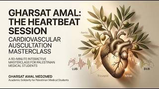
In this recorded lecture, Dr. Hassan Bencheqroun (MD, FCCP, MBA) provides a step‑by‑step instructional guide to cardiac auscultation for medical trainees. He begins by reviewing stethoscope mechanics—using the bell for low‑frequency sounds and the diaphragm for high‑frequency sounds—and then outlines the four primary valve auscultation zones (pulmonary, aortic, tricuspid, mitral) with precise intercostal space and sternal border landmarks, plus Erb’s point as a useful supplemental site. Dr. Bencheqroun explains the normal heart sounds S1 (closure of the atrioventricular valves) and S2 (closure of the semilunar valves), describing their physiological basis and the characteristic “lub‑dub” cadence. He introduces the additional sounds S3 and S4, detailing their timing relative to S1/S2, typical etiologies (e.g., rapid ventricular filling, stiff ventricle, heart failure), and the fact that they are often difficult to hear for beginners. The lecture then shifts to a practical, four‑step algorithm: (1) palpate a peripheral pulse (carotid or femoral) to anchor timing, (2) locate the cardiac apex (5th intercostal space, mid‑clavicular line) for S1 and the base (2nd intercostal spaces, right/left sternal borders) for S2, (3) coordinate the pulse upstroke with S1 and use kinesthetic learning (clapping, finger tapping) to differentiate S1 from S2, and (4) apply this framework to identify systole (S1‑S2) and diastole (S2‑S1). He emphasizes that the apex may shift laterally in cardiomegaly, so clinicians should slide the stethoscope along the 5th intercostal space to locate displaced apexes. The session concludes with a homework assignment to practice locating auscultation points, correlating sounds with the pulse, and distinguishing normal from abnormal heart sounds, followed by well‑wishes for Ramadan and a brief cultural sign‑off.
Chapters
Perfect. So several of you have asked me, you know, the labs that you were supposed to be learning how to auscultate, cardiac auscultation, were missed by some of you. And so today we're going to be talking about cardiac auscultation. And the cardiac auscultation is let's go to content only. Okay, I hope you can see my screen. All right, let's start first by— so when it comes to cardiac auscultation, we are going to be discussing using our stethoscope. So when it comes to auscultation, we can use the bell for low-pitched sounds, and we can use the diaphragm for high-pitched sounds. What does that mean? Well, the high-pitched sounds, it's like a child screaming. It's very high. And low-pitched sounds is like a bong of a church bell. So you can flip the stethoscope. Some stethoscopes have the ability to do that and some don't. What do we do with that stethoscope now that we have it? Well, the first thing that we do is there are 4 valve areas. The first one is the pulmonary area, and that's in the second intercostal space. Left sternal border. Then you have the aortic area. It's in the second intercostal space in the right sternal border. After that, we have the tricuspid area, and that's in the fifth intercostal space in the left sternal border. And then we have the mitral area in the fifth intercostal space in the midclavicular line. So, it's very important that we realize that these are the areas where you put your stethoscope in order to hear the heart sounds, the additional heart sounds, or if there are any murmurs. Some people also talk about the Erb's point, which is the third intercostal space, and that will give you, on the left sternal border, the ability to hear most heart sounds. That's S1, S2, S3, and S4. I hope this is clear, and that gives us where are we going to be auscultating the heart sounds. Now, when it comes to hearing those heart sounds, we are going to be discussing this. The normal heart sounds that you listen to with the stethoscope are S1, S2. Now, S1 is actually the closing of the atrioventricular valves or the AV valves, and that means it is the start of the systolic contraction of the ventricles. So as soon as you start doing listening to your heart squeezing blood out, it needs to go into the pulmonary and the aorta. It cannot go back from the ventricle to the atrium. So the valves between the atrium and the ventricle close, and that snap is what S1 is. S2 is actually the semilunar valves, the valves. It's once the systolic contraction is complete Once the systolic contraction is complete, what that means is it is preventing blood which has been already propelled forward into the aorta and into the pulmonary artery from going back into the ventricles. Those valves, once you leave from one chamber to the other, the valves close behind you. So you don't go back. So, when we have the contraction of the ventricles, the AV valves close. When the ventricles have finished contracting and the blood has left the heart, the semilunar valve, which is the pulmonary and the aortic valves, close, and that's S2. Okay? And when we talk about this, it's gonna prevent it from flowing back. And the way that it sounds is literally lub dub, lub dub, lub dub. Okay? So that will allow us to understand S1 and S2. What are third heart sound, S3, and the fourth heart sound, S4? S3 usually happens after S2, and it's not too far, so sometimes we get a little, "I can't hear it." Trust me, you will not hear it the first time somebody tells you there is an S3. You won't. Feel okay with that. It's gonna take a long time for your ear to become accustomed to an added sound, and it usually happens as a /dədət/, meaning it's so, so, so close to And you can hear that it's very difficult to hear that duplicate sound the second time around. So, what is the cause of S3? It's usually when you have the ventricle filling up so quickly that the chordae tendineae, or those cords in the ventricles attaching The valves are snapping almost like a guitar string. And it's normal in young patients sometimes because their heart is so quick that it fills up very quickly. But it's always abnormal in older patients where the heart is stiff and those chordae tendineae string snaps quickly. And it's usually S3. It's usually heart failure. Now, let's talk about the next one, which is the fourth heart sound. When we're talking about the fourth heart sound, meaning S4, S4 happens before S1. And you're probably gonna ask, well, hold on a second, I have S1 and S2. Isn't S3, that is after the S2, before the next S1? So how do I know? This is all in training your ear. You must be patient with it. You're gonna get it one day. You're not gonna get it the first day, and you should be patient with your ears. So the S4 is always abnormal. It's quite rare. And it's usually because the ventricle is very thick. And when there is a drop in that blood into the ventricle, it hits a very stiff, thick ventricle. And in that stiff, thick ventricle, and it drops, that turbulent flow is what you hear. and it usually hears like, "Lub dub, lub dub, lub dub." So you can hear that it happens before the S1. That's what makes the auscultation of the heart sounds. Now, So far, I want to make sure that everybody is following. Nothing in the chat. Give me a reaction. So far, so good. How are we doing with these sounds? Do you have an idea where to put the stethoscope now? Okay, hearing none and seeing no chat. No reaction. I'm going to follow. This is recorded anyways, so you can get back to it. Now we're going to go to here, and what I want here to be is for us to start identifying the heart. If you think that you need advanced clinical technology, think again. Take your hand off of the mouse and put your 2 fingers on the pulse. Check the pulse. That will anchor you. And this is our framework. Our framework is going to be Let's take a look at the pulse. But before we do that, what we're going to do is we're going to feel the pulse, then locate where is it that we are listening to the heart sounds. And then we're going to try to coordinate our pulse with those heart sounds and identify which valves. That timing allows us to know which valves are opening and which valves are closing. So, let's go ahead and identify the pulse. Carotid, right or left, or femoral. And looking at the diagram, it shows the conceptual foundation. Your first sound that you hear, S1 or lub, is the sound of the AV valves snapping shut. That's the core memory right there. Squeeze the heart means that we don't want the blood that just made it to the ventricle to go back into the atrium. So the AV valves, which means the mitral and the tricuspid, squeeze shut. That is the start of systole. Then we have the S2, and I'm repeating here, but this is because this is what the conceptual foundation is. The S2 is the semilunar valve closing, which are the aortic and the pulmonic valve. We do not want the blood that just flew, flowed from the ventricle to the pulmonary artery and the aortic the aorta to flow back into the ventricles. So, the semilunar valves close. That is the stop of systole. So, that's why this is important to understand the behavior of these valves as it explains the rhythm and the biological noise. Again, S1, lub, squeeze, start. Of the systole. S2 is dub, and it's the semilunar valves, and it's a squeeze, but it stops the systole. Okay? Now, when we look at this diagram and you try to coordinate it with your pulse, you will find S1 and S2 is defining the systole, and that's going to feel like the pulse. Another thing you can do is what we call kinesthetic learning. You have one hand that is always the one that moves and the other one that is stopped. You clap either to see where the S1 is or where the S2 is. And if you were to put the stethoscope in your ears and your fingers are on your pulse, you can coordinate S1 with the upper upstroke of the arterial pulse. And then you can start to clap in order to match the S1 versus the S2. That is the best way to identify S1 and S2 in order for you now to start paying attention to an S3 or an S4. The location is similar. The apex is what we have talked about before, which is at the bottom, and in the apex, you can hear the S1, and at the base, you can hear the S2. We can go back to the location map after we're done with this in order for us to realize how and where to look at this. I hope that this was a little bit explanatory. So these are what we discussed today, the 4-step algorithm that I want you to— you need to do the feel your pulse that is corresponding to the upstroke or S1, feel the location at the apex S1, S1 is louder and S2 is louder at the base. Correspond to the valves. The AV valves are S1. The semilunar valves are S2. And then the timing. S1 to S2 is the systole. S2 to S1 is the diastole. And then correspond them to the valves as we talked about. The AV valves are mitral and tricuspid, and semilunar valves are the aortic and the pulmonic. And then the timing. And that should help you in high-pressured environment when you don't have time. You would have trained in it already when nothing was happening. So that is your homework, is I want you to go home and start to look into these very important location sounds, correspond them to the pulse, and then identify where is S1 and S2 and where is S2 to S1. Where is the systole and where is the diastole? And what we're going to lastly take one last time is to take a look at the auscultation areas. And the auscultation areas are Right here. So the apex area is the 5th intercostal space midclavicular line. That's the mitral area. And the base area is the pulmonic and the aortic area. That's where you need to move your stethoscope to hear S1 and S2 better. One last thing I will share with you. You have your heart has a heart failure, which means that it is bigger than it should, dilated. The heart will not have the same anatomic areas, especially the apex. The apex area tends to move to the side. So, when you cannot hear it here, start to move your stethoscope along the 5th intercostal space towards the left side or the axilla, you may find that the apex is actually moving in that area because the heart is growing bigger. And hopefully with this, you have learned the principles of auscultation of the heart sounds and how to coordinate them with the pulse and the timing in order for you to start detecting what is abnormal. What's normal is the first thing to do before you start to hear the abnormal heart sounds. Is there any question at the moment? Have I missed anything today? Very good. I'm going to wish you a fantastic day. I sincerely, sincerely hope and wish for peace, for serenity, and for God and Allah to accept your Ramadan fasting. And for anybody else who is struggling, Allah irham shuhada al-muslimin. And I hope that this helps you even a little bit to feel supported. See you, inshallah, next week.